


Introduction
MicroRNAs (miRNAs) are a class of small, non-coding, and highly conserved RNAs that are ~20-22 nucleotides in length and control many biological functions through the posttranscriptional and translational regulation of mRNA. Binding to the 3′untranslated region (3′UTR) of the target, miRNAs adversely affect their expression, leading to the degradation of target genes’ transcription or translation inhibition [1,2]. miRNAs are transcribed as long primary transcripts from the genome (pri-miRNA) and are then cleaved by the endonuclease enzyme Drosha and the cofactor DGCR8 (DiGeorge syndrome critical region gene 8) in the nucleus [3]. Pre-miRNA, a stem-loop structure of approximately 70-100 nucleotides, is transferred to the cytoplasm by exportin 5 and processed into a double-stranded mature miRNA of approximately 20-22 nucleotides in length through cleavage by Dicer [4,5]. The guide strand of this mature miRNA, which binds to target genes, is associated with the RNA-induced silencing complex (RISC), thus contributing to decreased protein expression [6,7]. This pathway (Drosha/DGCR8) is the canonical pathway for miRNA biosynthesis [8]. In the non-canonical pathway, miRNAs are generated without processing by Drosha/DGCR8 but still require Dicer for maturation, or they are transferred directly from Drosha/DGCR8 to the RISC, and Dicer is not involved [9,10]. miRNAs can regulate multiple target genes simultaneously and are therefore involved in many vital cellular functions, such as the regulation of cell proliferation and differentiation, homeostasis, angiogenesis, development, the immune system, apoptosis, and hormone biosynthesis and secretion [11-15]. miRNA expression is tissue-specific and closely related to tissue function. This feature has also been observed in ovaries [16,17]. The ovary is a complex organ that contains oocytes and somatic cells such as granulosa, theca, and cumulus cells (CCs), which have specific miRNA profiles. Many miRNAs play important roles in the development of follicles, ovulation, and other developmental processes in the ovary [18]. Therefore, miRNAs can be attributed to various types of ovarian disorders and diseases, including polycystic ovary syndrome (PCOS) [19,20]. In patients with PCOS, abnormal expression of miRNAs in serum, follicular fluid, and granulosa cells has been detected, which is reportedly related to anti-apoptotic function and inconclusive testosterone response [17,21].
PCOS is the leading endocrine disorder among women of reproductive age worldwide. The frequency of PCOS is widely dependent on ethnicity, physical characteristics, and diagnostic factors [22,23]. The prevalence of PCOS is calculated as 15% by Rotterdam criteria, reaching 30% in obese and overweight women [24,25]. This disorder is diagnosed based on two of the following three criteria: menstrual irregularity (oligo-ovulation or anovulation), hyperandrogenism (clinical or biochemical), and polycystic ovarian morphology. PCOS is often associated with infertility, obesity, insulin resistance, and dyslipidemia [26-28]. This complex disorder is caused by a combination of environmental and genetic factors [29]. Ethnic diversity in patients with PCOS is strongly associated with their genetic background [30,31]. PCOS is one of the most common endocrine disorders in women and the most common cause of infertility due to lack of ovulation [32]. Moreover, miRNAs play a pivotal role in the posttranscriptional regulation of gene expression and signaling pathways. Therefore, we reviewed recent findings on the expression profiles of exosomal and non-exosomal miRNAs in serum and ovarian follicular fluid and their potential role in ovarian development and PCOS. A better understanding of their functions at different stages of ovarian development can shed new light on women’s reproductive health and the prediction and prevention of various ovarian disorders and infertility.
miRNA and ovarian development
Similar to other human tissues and organs, miRNAs are expressed in ovarian tissue (Fig. 1). These molecules play important roles in ovarian follicular development, including follicular growth, cell proliferation, atresia, and ovulation [33]. Ovarian development begins in the early embryonic period; however, follicular growth and ovulation, followed by luteal tissue development and cell death, continue throughout life. Targeted knockout of argonaute (AGO2) and Dicer in specific tissues of mouse models has revealed the valuable role of miRNAs in ovarian development and the function of various proteins involved in these pathways [34-36]. Bioinformatic and gene ontology analyses have shown that miRNA-related target genes in the ovary are involved in cell cycle regulation, growth, proliferation, apoptosis, endocrine disorders, and ovarian function [37,38]. miRNAs also play a role in the regulation of ovarian follicle growth and are involved in certain stages of ovarian follicular development, ovulation, luteal function, and period.
1. miRNAs in the follicular developmental phase
The miR-17-92 cluster, miR-124, and Let-7 are involved in the growth and development phase of primordial cells; miR-224 in the secondary primordial phase; miR-21, miR-132, miR-212, miR-513a-3p, and miR-125b in the antral phase; and miR-21 and miR-224 in ovulation [18,39]. miR-199a-3p, miR-145, and miR-31 are overexpressed in the follicular phase and substantially decreased during the transition to the luteal-follicular phase [40,41]. miRNAs also play a role in gonadal differentiation. miR-124 is one of the miRNAs that target SOX9 in undifferentiated gonads. Increased expression of SOX9 leads to testicle formation, whereas low levels of SOX9 result in ovarian formation [42]. miR-17-92 cluster miRNAs, except for miR-17-3p, are highly expressed in primordial germ cells (PGCs), and several studies have demonstrated the importance of this cluster in primary ovarian primordial follicle development [43]. For example, miR-143 inhibits primordial follicle formation by reducing the expression of cyclin-dependent kinases (CDKs) 4 and 6 and cyclin B1 in pre-granulosa cells [44]. In mouse ovaries, miR-181a inhibits granulosa cell proliferation by targeting activin receptor IIA [45]. Moreno et al. [46] analyzed the miRNA profiles in the follicular fluid of oocytes in metaphase II (MII), metaphase I (MI), and germinal vesicles (GV). Comparing GV and MII, 13 differentially expressed miRNAs were found, of which miR-451 and miR-563 upregulated in follicular fluid of oocytes in GV and MII stages, respectively. In addition, a comparison of miRNA profiles of the follicular fluid of MII and MI showed that miR-451 was highly expressed in MI and miR-574-5p in MII. Given these findings, they suggested that miR-451 functions as a predictor of the early stages of oocyte maturation. The potential gene targets of these miRNAs are related to the kinase, calcium, and mitogen-activated protein kinase (MAPK) signaling pathways, which are involved in the production of follicle-stimulating hormone and luteinizing hormone (LH) and, in turn, can influence the proper timing of meiosis and oocyte maturation [46]. Studies using mouse models have suggested that the Lin28/Let-7 system is a functional miRNA system involved in ovarian development. Lin28 is an RNA-binding protein that downregulates Let-7. Recent studies have shown that this pathway is involved in the proliferation of PGCs. Although the presence of Let-7 is essential for PGC proliferation and normal mature ovary function during ovarian development and its early stages, the expression of Lin28 is necessary to reduce the expression of Let-7 [47]. A new isoform of Dicer has been identified in recent investigations on mouse and rat oocytes, revealing that Lin28/ Let-7 may play a vital role in oocyte maturation, fertilization, and embryonic development in other species. Let-7b is also expressed in granulosa and CCs of the ovary and is involved in folliculogenesis and ovarian follicle development through regulating the transforming growth factor (TGF)-β signaling pathway [48].
2. miRNAs in the luteal phase
Let-7b, miR -17-5p, miR-122, miR-125, and miR-378 are involved in corpus luteum formation [18,39]. In addition, miR-503, miR-21, and miR-142-3p are generally expressed at lower levels in the follicular stages, and their expression is considerably higher in luteal tissues [40,41]. miRNAs also play a role in the post-ovulation phase, when follicular atresia is caused by granulosa cell apoptosis. MiR-34s activates cell apoptosis and growth arrest through p53 activation and the CDK inhibitor p21 [49]. miR-21 is also highly induced in mouse granulosa cells by corpus LH, and suppression of miR-21 activity in vitro causes granulosa cell apoptosis [50].
Non-exosomal miRNA in PCOS
miRNAs exhibit tissue-specific expression patterns in various organs and systems. Data from various studies have shown that changes in serum levels of miRNAs are valuable and important tools for the diagnosis, tracking, prognosis, and treatment of numerous diseases. This is mostly due to their unique characteristics, such as stability, resistance to nuclease degradation, simple detection, and the potential to be disease-specific [51]. The mechanism by which miR-NAs are transferred from inside the cell to the extracellular environment, such as serum, is accomplished through two main pathways: 1) active transfer via extracellular vesicles, such as exosomes and 2) transfer as part of a protein-miRNA complex; they can also leak from damaged cells [52]. Table 1 shows the various miRNAs and their potential functions in different parts of ovarian tissue.
1. Serum
Several studies have shown that changes occur in the serum levels of different miRNAs in patients with PCOS (Fig. 2). Since serum levels are easily detectable, they can be used as important non-invasive biomarkers for early diagnosis of PCOS [53]. Long et al. [54] observed that increased serum levels of miR-222, miR-146a, and miR-30 might play a role in the pathogenesis of PCOS. Bioinformatics analysis showed that the target genes of these miRNAs were involved in metastasis, cell cycle, apoptosis, and endocrine pathways [54]. In patients with PCOS, the level of testosterone increases, and insulin contributes to this by upregulating the synthesis and accumulation of androgens in the adipose tissue [55]. In addition, miR-132, miR-320, miR-520c-3p, miR-24, and miR-222 regulate estradiol concentrations, while miR-24, miR-193b, and miR-483-5p regulate progesterone concentrations in patients with PCOS [56]. Studies have demonstrated that miR-222 is directly correlated with serum insulin levels, and miR-146a is inversely associated with serum testosterone [57]. Changes in the serum levels of miR-222, miR-146a, and miR-30 have been introduced as new biomarkers for the diagnosis [42]. miRNAs are also involved in the pathogenesis of PCOS through metabolic processes. For example, miR-93 is overexpressed in PCOS and associated with decreased glucose transporter 4 (GLUT4) expression and increased insulin resistance [58]. Additionally, miRNA-21, miRNA-27b, miRNA-103, and miRNA-155 play important roles in metabolic processes and are affected by obesity and circulating androgen concentrations in patients with PCOS [59]. miR-103 is one of the miRNAs involved in insulin resistance and obesity and plays a role in regulating testosterone levels and the occurrence of PCOS. Additionally, it increases in serum and is therefore easily detectable; making its serum level be considered as a biomarker for PCOS, especially in obese women [60]. Research conducted on miR-23a and miR-23b showed a positive effect of obesity on the serum expression of miR-23b, an adverse effect of testosterone on the expression of miR-23a, and decreased expression of these two miR-NAs in the serum of patients with PCOS compared to healthy women. Although, miR-23a is a more effective option concerning new biomarkers for PCOS diagnosis [61]. miRNA-93 and miRNA-223 were significantly upregulated in the blood of patients with PCOS compared to controls. There is no association between miRNA-93 or miRNA-223 with insulin, homeostatic model assessment for insulin resistance, homeostasis model assessment of beta cell function, or testosterone levels in both groups. Plasma levels of miR-93 can differentiate patients with PCOS from healthy subjects with greater sensitivity and specificity than miR-223. Thus, circulating miR-93 could serve as a biomarker for PCOS [62]. miR-320 is one of the other serum miRNAs related to the PCOS phenotype, whose serum level decreases compared to the normal group, and can also be used as a non-invasive diagnostic biomarker [63,64]. In addition, miR-320 is downregulated in follicular fluid [56] and ovarian tissue [65]. Endothelin (ET-1) is a bioactive peptide produced by endothelial cells that participates in tumor growth by enhancing cell mitosis [66]. miR-320 is involved in follicular maturation by targeting the ET-1 gene, and a decrease in miR-320 can lead to the progression of the PCOS phenotype in affected women [46]. Changes in miR-NA-21 expression were observed in both ovarian tissue and serum. Increased expression of miR-21 acts as an inhibitor of the serine/threonine protein kinase LATS1 and stimulates the abnormal growth of secondary follicles by affecting the Hippo signaling pathway and increasing the level of cellular communication network protein, which is involved in the further growth, viability, and proliferation of cells [67]. miRNA-6767-5p, involved in the cell cycle and immune system processes, plays an important role in ovarian development [49]. In a study conducted on Korean women with PCOS, Sang et al. [56] observed that the serum level of miR-6767-5p was inversely correlated with fasting glucose levels and directly correlated with the number of menstrual periods per year. As hyperandrogenemia is the main characteristic of PCOS, miR-6767-5p may be of great importance in the pathogenesis and metabolic manifestations of PCOS. They suggested that serum miR-6767-5p may serve as a new molecular marker for the diagnosis of PCOS in Korean women [68].
2. Ovarian tissue
In a study of the expression profiles of miRNAs in the ovarian tissue of women with PCOS, Baley and Li [35] observed that some miRNAs, such as miR-146a, miR-22, miR-132, miR-200c, miR-141, and miR-21, showed significantly increased expression. Interestingly, most differentially expressed miRNAs in the ovarian tissue of patients with PCOS were expressed at normal levels in serum. Therefore, all miRNAs in the PCOS ovarian tissue are released into the blood. MiR-142, miR-33b, and miR-423 are other miRNAs involved in PCOS that contribute to the initiation and progression of the PCOS phenotype by targeting Is transforming growth factor beta receptor 1 and mothers against decapentaplegic homolog 7 (SMAD7) and inhibiting the TGFB signaling pathway [69]. Bioinformatics analyses have revealed that dysregulation of miR-15b and miR-497, which are miRNAs involved in the insulin signaling pathway, contributes to PCOS pathogenesis [70]. miR-320 also regulates the expression of insulin receptor substrate (IRS)-1 and the extracellular signal-regulated kinase (ERK) 1/2 activity in ovarian tissue, which are related to insulin resistance in PCOS. Therefore, understanding the mechanism of action of miRNAs in PCOS patients with insulin resistance presents a theoretical basis for the diagnosis and treatment of this condition [65,71]. The inhibition of miR-483 expression has been observed in the ovarian cortex of patients with PCOS. miR-483 is downregulated in PCOS and prevents granulosa cell proliferation by targeting insulinlike growth factor (IGF)-1 [72]. miR-33b-5p is overexpressed in the ovarian tissues of insulin-resistant mice with PCOS and thus may play an important role in the development of insulin resistance in patients with PCOS. Additionally, miR-33b-p inhibited the production of GLUT4 by targeting high-mobility group AT-hook 2 [73]. Another study identified 29 differentially expressed miRNAs in patients with PCOS. In these patients, miR-382-5p correlated with age and free androgen index (FAI), miR-199b-5p with anti-müllerian hormone (AMH), and miR-93-3p with C-reactive protein. In healthy controls, miR-127-3p, miR-382-5p, and miR-425-3p correlate with fertilization rate, miR-127-3p with insulin resistance, and miR-381-3p with FAI [74].
3. Follicular fluid
Ovarian follicular fluid plays an important role throughout follicular development. Being a complex mixture of hormones, growth factors, anti-apoptotic factors, proteins, and miRNAs secreted from the granulosa and theca cells, it provides a vital microenvironment for oocyte development and maturation of oocytes [75]. Scalici et al. [76] reported that the expression of Let-7b and miR-140 is significantly decreased, whereas the expression of miR-30a is increased in the follicular fluid of patients with PCOS. The results showed that reduced expression of Let-7b and miR-140 contributed to the progression of the PCOS phenotype. In addition, the change in the expression of estrogen receptor in PCOS was attributed to reduced expression of miR-140 in the follicular fluid [76].
In Roth et al. [77] study, five overexpressed miRNAs (hsamiR-9, 18b, 32, 34c, and 135a) were found in follicular fluid, and three of their potential targets were associated with the PCOS phenotype (IRS-2, synaptotagmin 1, and interleukin 8). These three genes are involved in carbohydrate metabolism, beta cell function, cell-cell communication, and steroid synthesis in the late follicular phase [77]. Some miRNAs are involved in the metabolism of hormones; for example, changes in the expression of some miRNAs involved in the metabolism of androgens have been observed in the follicular fluid of patients with PCOS. Among these, miR-151 was inversely correlated with free testosterone, whereas miR-29a, miR-320, and miR-518 were directly correlated with testosterone levels. miR-132, miR-135, and miR-146a inhibit progesterone and testosterone secretion, whereas miR-9, miR-18b, and miR-155 inhibit testosterone secretion [75]. In this regard, Naji et al. [78] observed that the intermediary role of (miR-93 and miR-21) miRNAs in granulosa cells and follicular fluid plays an important role as an androgen-responsive factor in the pathogenesis of PCOS in hyperandrogenism.
4. Granulosa cells
miR-145 plays a prominent role in granulosa cell proliferation, follicle selection, and folliculogenesis. And miR-182 plays an essential role in the appropriate functioning of granulosa cells by regulating proliferation, apoptosis, and steroidogenesis. Thus, aberrant expression of miR-182 (in follicular fluid) and miR-145 (in granulosa cells) may contribute substantially to follicular developmental disorders in PCOS [78]. Overexpression of miR-145 inhibits the mitogenactivated protein kinases (MAKP)/ERK signaling pathway in granulosa cells. These findings have also shown that miR-145 can suppress cell proliferation and that the mechanism involved is related to the suppression of the expression of IRS-1, which leads to the inhibition of the MAKP/ERK signaling pathways. In addition, high insulin concentrations decreased miR-145 expression, regulated IRS-1 protein expression, and promoted cell proliferation [79]. The expression of miR-483-5p in PCOS granulosa cells also increases and regulates the MAPK3 pathway [80]. Dihydrotestosterone (DHT) increases granulosa cell apoptosis and, at high concentrations, inhibits miR-182 expression. miR-182 inhibits granulosa cell apoptosis by targeting SMAD7, as well [81]. miRNAs are involved in the regulation of various pathways, biological functions, and cellular components in PCOS. Xue et al. [82] reported increased expression of miR27a-3p in PCOS mouse ovaries, and its function was investigated in mouse primary granulosa cells. They confirmed that the expression of miR-27a-3p in mouse granulosa cells increases in the pre-antral follicle stage and that the cyclic AMP response element binding protein 1 is a target gene for miR-27a-3p, which prevents the expression of the downstream factor CYP19A1 (cytochrome P450 family 19 subfamily A member 1). The imbalance in androgen and estradiol levels under the influence of miR-27a-3p and its function in promoting granulosa cell apoptosis may be involved in the pathophysiology of PCOS [83]. In addition, Zhang et al. [84] observed that a reduction in miR-320a impairs steroidogenesis in cumulus granulosa cells by dysregulating the function of the bone transcription factor RUNX2. CCs are the main site of estrogen synthesis. Bidirectional communication between CCs and follicular fluid is essential for ovarian steroidogenesis. miR-320a is markedly decreased in the primary CCs of patients with PCOS, which results in a sharp drop in estrogen levels in CCs. IGF-1 regulates the expression of miR-320a in CCs, and miR-320a can promote steroidogenesis in CCs by decreasing the expression of CYP11A1 and CYP19A1 by directly targeting the 3’UTR of RUNX. Therefore, deregulation of the miR-320a/RUNX2/CYP11A1 (CYP19A1) cascade plays an important role in the development of estrogen deficiency in human CCs. Androgens influence the growth, health, and survival of follicles and the pathophysiology of PCOS. Investigations have revealed that miR-93 and miR-21 are increased in the granulosa cells of hyperandrogenic PCOS patients compared to normoandrogenic patients. Free testosterone and FAI were directly correlated with miR-93 and miR-21 in PCOS Granulosa cells. Thus, as miR-93 and miR-21 are widely known androgen-responsive factors, they may be involved in follicular dysfunction during the pathogenesis of PCOS in hyperandrogenism [85]. Transfecting miR-509-3p mimic in KGN cells and studying E2 production confirmed that miR-509-3p improves estradiol secretion by inhibiting MAP3K8 expression. These results help to define the pathogenesis of anovulation in PCOS, particularly the regulation of estradiol secretion [86]. miR-93 expression is upregulated in granulosa cells of PCOS patients and inhibits cyclin-dependent kinase inhibitor 1A (CDKN1A) in PCOS granulosa cells. MiR-93 causes granulosa cell proliferation and G1/S transition. Therefore, miR-93 promotes ovarian granulosa cell proliferation through targeting CDKN1A in PCOS [21,87].
5. Theca cells
Ovarian theca cells play a vital role in ovarian follicular development and hormone secretion [88]. Differential expression of some miRNAs has been reported in theca cells, which may regulate various signaling pathways including meiosis, WNT, TGF-β, and MAPK [89]. A study of miRNAs in atretic follicles compared to healthy cattle follicles revealed that miR-150, miR-409a, miR-142-5p, miR-222, miR-155, and miR-199a-5p were more highly expressed in the theca than in the granulosa cells of atretic follicles [90]. In a rat POCS model induced by DHT, most of the miRNAs that stimulated cyst formation were expressed in follicular theca cells [91,92]. Hossain et al. [93] reported the upregulation of miR-222 in theca cells of PCOS samples; however, androgens can target P27/kip1(a cyclin-CDK inhibitor) to regulate the proliferation of theca cells, leading to miR-222 suppression. In vitro studies have confirmed that PCOS theca cells secrete higher androgen levels than normal cells [94]. In addition, miR-103a-3p and miR-376a-3p positively correlated with androgen levels in PCOS [95]. Therefore, miRNAs may be implicated in pathways involved in insulin signaling, steroid biosynthesis, endothelial regulation, and folliculogenesis.
Exosomal miRNA in PCOS
Exosomes are a heterogeneous group of extracellular microspheres with diameters of approximately 4-100 nm [96]. Exosomes exist in different types of mammalian cells in the form of multivesicular bodies in the intracellular space and are secreted out of the cell or into biological fluids such as joint fluid, amniotic fluid, plasma, cerebrospinal fluid, urine, saliva, breast milk, alveolar fluid, follicular fluid, and bile through integration with the plasma membrane [97,98]. Exosomes may have different surface markers depending on their cellular origin and physiological state. They contain a variety of cell/tissue-specific proteins, RNAs, lipids, and biological biomolecules that facilitate cell communication with the surrounding environment and have a significant impact on biological and pathological events [99]. AGO2 and some RNA-binding proteins such as hnRNPA2B [100] and Y-box1 [101] are involved in the regulation of miRNA loading into exosomes. During the formation of multivesicular bodies in the cytoplasm, AGO2 binds to the exosome marker protein CD63, leading to the loading of miRNAs into exosomes [102]. Table 2 displays the exosomal miRNAs compared to non-exosomal miRNAs in patients with PCOS.
1. Serum
Che et al. [103] exposed endometrial cancer cell lines to serum exosomes isolated from patients with PCOS and found that the rates of migration and invasion of cancer cells increased in vitro. Further investigations showed that levels of miR-27-5p in the serum exosomes of patients with PCOS were significantly increased. They observed that exosomal miR-27-5p stimulated the migration and invasion of endometrial cells by targeting SMAD4 and reducing its expression. Therefore, miR-27-5p plays an important role in the esophageal carcinoma progression of endometrial cancer in PCOS patients [103]. Furthermore, the interaction between insulin resistance and obesity in PCOS may be related to the regulation of exosome-secreted miRNAs. An insulin resistance-based model of PCOS was established in rats with dehydroepiandrosterone (DHEA) on a high-fat diet (HFD). Expression of miR-20b-5p and miR-106a-5p was detected in serum exosomes. Overexpression of serum exosomal miR-20b-5p and miR-106a-5p inhibits adipocyte differentiation in 3T3-L1 cells in a mouse model of PCOS with IR. In DHEA+HFD serum-derived exosomes, miR-20b-5p and miR-106a-5p levels were significantly decreased. Overexpression of miR-20b-5p and miR-106a-5p decreased the expression of genes related to adipose differentiation and triglyceride content in 3T3-L1 cells of mice with liver steatosis. Serum-derived exosomal miR-20b-5p and miR-106a-5p inhibit adipocyte differentiation during PCOS with IR and therefore have the potential to serve as new therapeutic targets [104]. Another study has investigated the expression profiles of exosomal miRNAs in PCOS and non-PCOS patients. Hsa-miR-1299, hsa-miR-145-5p, hsa-miR-6818-5p, and hsa-miR-192-5p were found to be differentially expressed in the serum exosomes of PCOS patients and therefore have the potential to be used as diagnostic biomarkers for PCOS [105]. Circulating exosomes in the PCOS plasma contain differentially expressed miRNAs. In patients with PCOS, the expression levels of miR-146a-5p and miR-126-3p increased, whereas those of miR-20b-5p, miR-106a-5p, and miR-18a-3p decreased. The differential expression of these miRNAs has been suggested to target various functions, including the MAPK signaling pathway, axon guidance, endocytosis, and tumorigenic pathways. Therefore, these exosomal miRNAs may be associated with PCOS [106].
2. Follicular fluid and other cells
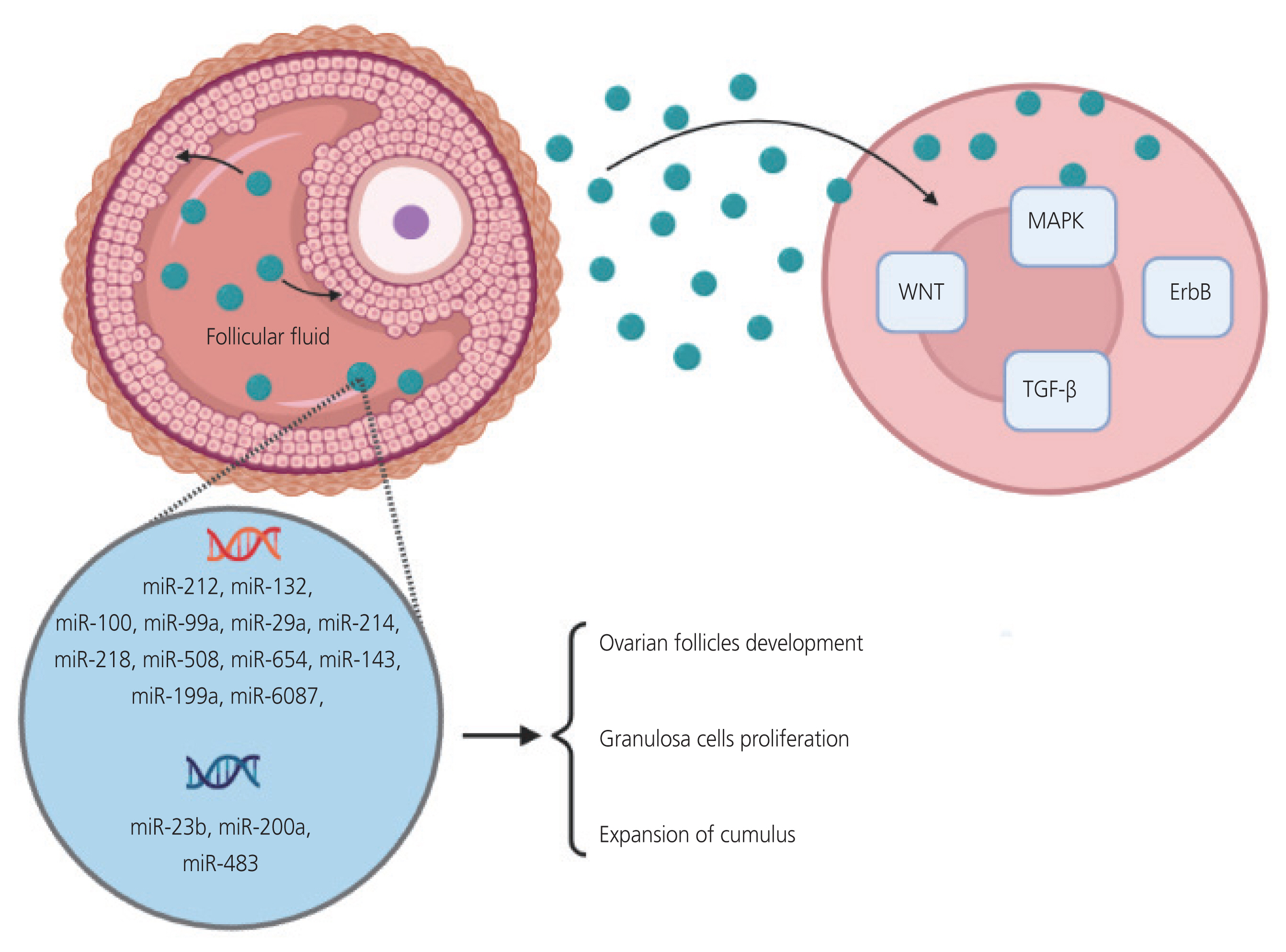
Various studies have shown that more than 200 different miRNAs exist either freely or inside human follicular fluid exosomes [107]. Fig. 3 shows some miRNAs in the follicular fluid exosomes. Developmental pathway analysis showed that the exosomal and non-exosomal miRNAs target genes are involved in MAPK, Wnt, FOXO, nuclear factor kappa B, Oxytocin, epidermal growth factor (ErbB), PI3K-Akt, and neurotrophin signaling pathways, which play role in follicular development, cell proliferation, and resumption of meiosis [108]. Li et al. [109] study showed that the S100-A9 protein in exosomes could activate NF-κB signaling pathways in granulosa cells, increase the production of inflammatory cytokines, and disrupt steroidogenesis. These findings indicate the importance of exosomes as extracellular mediators in the pathophysiology of PCOS [109]. Increased expression of miR-29a, miR-99a, miR-100, miR-132, miR-212, miR-214, miR-218, miR-508-3p, and miR-654-3p, all molecular regulators of ovarian follicle development, was observed in exosomes isolated from the follicular fluid. Some of these miRNAs or their host genes are expressed in granulosa or CCs. However, they also play a role in targeting genes within follicular cells. Most of the upregulated miRNAs are involved in the regulation of WNT, MAPK, ErbB, and TGF-β signaling pathways, and can contribute to the development of ovarian follicles or stimulate the proliferation of granulosa cells and expansion of cumulus. They can also start the resumption of meiosis by negatively regulating genes that encode follicle maturation-inhibiting factors [110]. Hu et al. [111] identified six exosomal miRNAs in the follicular fluid (miR-6087, miR-199a-5p, miR143-3p, miR-483-5p, miR-23b-3p, and miR-200a-3p) related to PCOS pathogenesis and predicted that these miRNAs were mainly related to amino acid biosynthesis; metabolism of glycine, serine, and threonine; glycosaminoglycan biosynthesis; monocarboxylic acid metabolism; and carbon metabolism. Yang et al. [112] introduced three exosomal miRs, miR-125b, miR-19b, and miR-222, found in the follicular fluid associated with PCOS pathogenesis.
Inhibition of apoptosis and increased senescence in granulosa cells can also be mediated by exosomal miRNAs. The expression of miR-424-5p significantly decreases in PCOS exosomes and primary granulosa cells. Exosomes enriched with miR-424-5p significantly increased granulosa cell senescence and suppressed cell proliferation. Consistent with the results obtained in cells transfected with miR-424-5p mimic, miR-424-5p caused senescence and inhibited cell proliferation. The cell division cycle 4 (CDCA4) was identified as a direct target of miR-424-5p. Overexpression of CDCA4 reverses the effects of exosomal miR-424-5p on granulosa cells, activates the Rb/E2F1 signaling pathway, induces cellular senescence in PCOS, and attenuates the disease [113]. However, exosomal miRNAs can induce opposite effects. For example, exosomes containing miR-143-3p derived from PCOS follicular fluid increased granulosa cell apoptosis by targeting bone morphogenetic protein receptor and blocking the Smad1/5/8 signaling pathway [114]. miR-18b-5p produced by follicular fluid-derived exosomes decreases phosphatase and tensin homolog expression and activates the the phosphatidylinositol-3-kinase/akt and the mammalian target of rapamycin (PI3K/Akt/mTOR) signaling pathway to improve PCOS [115].
Significance of miRNA in therapy
miRNAs are among the most significant RNA groups in clinical research. Because a specific miRNA may be involved in different biological pathways disrupted in a disease, it can be very useful from a therapeutic perspective, especially if the disease is not associated with a genetic defect. In this regard, bioinformatics software used to identify miRNA binding sites, target genes, and the biological pathways involved, along with laboratory in vitro and in vivo research models, have greatly assisted in accelerating the identification of miR-NAs in clinical medicine [116]. In healthy individuals, a large proportion of ovarian follicles undergo apoptosis and die during their reproductive life through a vital process known as follicular atresia [117]. Follicular atresia begins with the apoptosis of follicular granulosa cells (granulosa cells), followed by the degeneration of other follicular components. Factors such as IGF-1 are follicle viability factors that control atresia [118]. Das et al. [119] showed that there was a significant variation in the apoptosis rate and proliferation of granulosa cells in patients with PCOS. They were the first to find that the level of active caspase-3, which is functionally required to initiate cell death, was significantly lower in the granulosa cells of anovulatory PCOS follicles than in those of normal ovulatory follicles. The expression of Bcl-XLong, an anti-apoptotic factor, increased approximately two-fold, while that of Bax, an apoptosis inducer, slightly decreased in the PCOS group. The expression of Ki-67 (an indicator of cell proliferation) was significantly higher in the PCOS group, indicating a significant imbalance between the rates of apoptosis and proliferation in patients with PCOS [119]. Studies have shown that miRNAs are involved in this pathway, and their regulation can be useful in disease treatment. miR-99a is one of the miRNAs found in granulosa cells, and its expression is significantly decreased in PCOS. This decrease was associated with an increase in the IGF-1R protein levels. IGF-1R belongs to the transmembrane receptor tyrosine kinase family and is activated by IGF-1 [120]. During follicular development, the increase in IGF-1R through the stimulation of tyrosine kinase pathways leads to an increase in proliferation and a decrease in apoptosis of granulosa cells in the first stage of folliculogenesis, which contributes to the pathogenesis of PCOS. Therefore, the re-regulation of miR-99a may be an effective method for reducing the adverse effects of IGF-1/IGF-1R system overactivation in PCOS [121]. miR-323-3p is another miRNA involved in the pathogenesis of PCOS that regulates cell apoptosis and steroidogenesis by targeting IGF-1. Studies have shown that the expression of miR-323 declines in CCs of PCOS patients [122]. CCs, a subclass of granulosa cells that surround the oocyte in the antral follicle, are the source of ovarian steroid metabolism and growth hormones and play an important role in oocyte maturation [123]. Apoptosis is one of the primary causes of PCOS [124]. Increased miR-323-3p expression in CCs stimulates cell proliferation and inhibits apoptosis. As previously mentioned, CYP19A is a critical enzyme in the production of ovarian estradiol, which increases in CCs in PCOS [125]. The expression level of this enzyme is indirectly controlled by miR-323; thus, the reduction of miR-323 in PCOS CCs leads to an increase in the level of IGF-1, followed by an increase in CYP19A, and a disruption in the completion of oocyte development. Thus, miR-323-3p is a novel and promising molecular target for ameliorating CC dysfunction in PCOS [105]. Programmed cell death 4 (PDCD4) is a key factor in apoptosis that suppresses protein translation by binding to eIF4A and inhibiting RNA helicase activity [126]. Available evidence demonstrates that the expression of PDCD4 increases in patients with PCOS and is involved in the pathogenesis of the disease [127]. In an investigation of 36 female C57BL/6 mice, Zhao et al. [128] observed high PDCD4 expression in mice with PCOS compared to that in controls. Delivery of exogenous miR-323-3p by adipose mesenchymal stem cell derived exosomes can suppress the expression of PDCD4. As the apoptosis of CCs increases in PCOS to induce premature follicular atresia, studies have suggested that PDCD4 contributes to premature apoptosis in PCOS CCs, and its pro-apoptotic effect can be eliminated by miR-323-3p. Therefore, miR-323-3p upregulation suppresses CCs apoptosis by targeting PDCD4 in PCOS, and the delivery of miR-323-3p via adipose-derived mesenchymal stem cell exosomes is an effective approach for regulating PCOS progression [128]. miR-135 is another miRNA found in granulosa cells and its expression is highly upregulated in PCOS. miR-135a is regulated by androgens at the transcriptional level. Increased androgen levels in PCOS lead to an increase in miR-135a, and miR-135a can suppress proliferation, enhance the DNA damage response, and induce apoptosis by directly binding to the vascular endothelial growth factor C gene. Consequently, the rate of apoptosis increases during the middle stage of the antrum, leading to cyst formation. Therefore, regulating miR-135a expression can be considered a new therapeutic goal for PCOS patients [129]. miR-335-5p expression is inversely correlated with the number of antral follicles, AMH and testosterone levels. Research has shown that miR-335 levels in the granulosa cells and follicular fluid are significantly decreased in patients with PCOS. Bioinformatics analyses have shown that SGK3 is the direct target of this miRNA, a member of the family of kinases involved in cell proliferation through the PI3K/AKT-mTOR signaling pathway [130,131]. In granulosa cells, SGK is localized in the cell nucleus and regulates cell proliferation and differentiation. In the early stages of folliculogenesis, a reduction in miR-335 levels leads to an increase in AMH levels and granulosa cell proliferation. AMHs prevent the premature depletion of ovarian follicles and regulate steroidogenesis in granulosa cells [132]. In contrast, the reduction of miR-335 expression in an SGK-dependent pathway leads to increased proliferation of granulosa cells, and delayed follicular atresia, and contributes to PCOS pathogenesis. These observations provide a new perspective on the function of granulosa cells in PCOS and suggest that miR-335-5p can serve as a new molecular target for improving dysfunctional granulosa cells [131]. Díaz et al. [133] investigated circulatory concentrations of miR-451a, miR-652-miR-451a, miR-652-3p, miR-106b-5p, and miR-206 in girls with PCOS treated with oral contraceptive or spironolactone-pioglitazone-metformin. They observed that the concentration of the studied miRNAs decreased significantly compared with that in the control group. These target genes are involved in energy homeostasis and cell-cycle regulation. This study demonstrated the capability of miR-451a to diagnose PCOS with 100% sensitivity [133].
Additionally, alterations in the circadian rhythm can trigger PCOS through oxidative stress and inflammation. Circadian rhythms can alter circulating exosomes and related miRNAs, leading to alterations in physiological conditions [134]. miR-NAs play an important role in maintaining homeostasis of the circadian system, and miR-132 and miR-219 are involved in regulating circadian rhythms in mammals by targeting clock genes. Furthermore, miR-494, miR-27b-3p, miR-155, and miR-142-3p are involved in regulating the clock gene, brain, and muscle Arnt-like protein 1 (BAML1) [135,136]. Changes in the expression of miR-192 and miR-194 not only affect the rhythm fluctuation of BMAL1 mRNA but also suppress the expression of the clock gene period (Per) [137]. Therefore, circadian rhythms can be modulated by targeting miRNAs to investigate their potential in PCOS treatment [134].
Conclusion
miRNAs that are free or enclosed in extracellular vesicles (exosomes) can affect various signaling pathways. Abnormal expression of miRNAs occurs in the granulosa cells, theca cells, adipose tissue, follicular fluid, serum, and peripheral blood leukocytes of women with PCOS. They regulate follicular growth and maturation, insulin signaling, glucose and lipid metabolism, and steroid hormone synthesis. Additionally, exosomes can protect miRNAs from degradation, making them stable and efficient diagnostic tools.
The use of miRNAs in the clinical management of PCOS is a dynamic part of research, and there are several potential functions of miRNAs in this context. miRNAs can be used clinically in various pathways, such as diagnostic and prognostic biomarkers, therapeutic targets, and drug delivery vehicles; therefore, as diagnostic and prognostic biomarkers of PCOS. Many studies have identified specific miRNAs that are dysregulated in individuals with PCOS, and their detection in the blood or other fluids may be useful for early diagnosis and monitoring of disease progression. Also, the expression of certain miRNAs has been found to be associated with insulin resistance, a common feature of PCOS. By measuring the levels of these miRNAs, it may be possible to predict the probability of increased IR-related complications, such as type 2 diabetes.
Although miRNAs are potentially promising diagnostic and therapeutic factors, their clinical applications in diagnosis and treatment are still under development. Concerning diagnosis, many of the identified miRNAs are still at the experimental level and require further investigation. Additionally, only a limited number of miRNAs have been approved by the Food and Drug Administration as therapeutic agents. Therefore, the application of miRNAs as diagnostic or therapeutic factors in medicine is in its early stages, and there is still a long way to go.


")


