The timing of adenomyosis diagnosis and its impact on pregnancy outcomes: a national population-based study
Article information

Abstract
Objective
Adenomyosis impacts pregnancy outcomes, although there is a lack of consensus regarding the actual effects. It is likely, however, that the severity of adenomyosis or ultrasound findings or timing of diagnosis can have different effects on adverse pregnancy outcomes (APOs).
Methods
In this study, we aimed to investigate the impact of the timing of adenomyosis diagnosis on pregnancy outcomes. Singleton pregnant women who delivered between 2017 and 2022 were analyzed based on the timing of adenomyosis diagnosis, using a national database. The final cohort was classified into three groups: 1) group 1, without adenomyosis; 2) group 2, those diagnosed with adenomyosis before pregnancy; and 3) group 3, those diagnosed with adenomyosis during pregnancy.
Results
A total of 1,226,475 cases were ultimately included in this study. Women with a diagnosis of adenomyosis had a significantly higher risk of APOs including hypertensive disorder during pregnancy (HDP), gestational diabetes mellitus (GDM), postpartum hemorrhage, placental abruption, preterm birth, and delivery of a small-for-gestational-age infant even after adjusting for covariates. In particular, concerning HDP, the risk was highest in group 3 (group 2: adjusted odds ratio [aOR], 1.15 vs. group 3: aOR, 1.36). However, the highest GDM risk was in group 2 (GDM; group 2: aOR, 1.24 vs. group 3: aOR, 1.04).
Conclusion
The increased risk of APO differed depending on the timing of adenomyosis diagnosis. Therefore, efforts for more careful monitoring and prevention of APOs may be necessary when such women become pregnant.
Introduction
Adenomyosis is a complex gynecological condition characterized by the presence of endometrial epithelial and stromal cells within the myometrium. Its prevalence is estimated to be between 20% and 35% [1,2]. This condition exhibits a wide range of anatomical and clinical variations, including differences in uterine size and symptoms, which can range from severe dysmenorrhea and heavy menstrual bleeding to being completely asymptomatic [3].
Adenomyosis can significantly impact pregnancy outcomes. Women with adenomyosis can face challenges in achieving pregnancy and have an increased risk of miscarriage. Additionally, adenomyosis has been associated with a higher likelihood of preterm birth, preeclampsia, and delivery of a small-for-gestational-age baby, as well as an elevated rate of delivery by cesarean section [4–6]. While the impact of adenomyosis on pregnancy can vary among individuals, it underscores the importance of comprehensive prenatal care and close collaboration between patients and healthcare providers to optimize maternal and fetal well-being.
However, assessing the severity of symptoms or interpreting ultrasound results involves subjectivity, which presents challenges for conducting research. Moreover, existing research on the effects of adenomyosis has several limitations that warrant consideration. The diagnostic criteria for adenomyosis vary among studies, using methods such as transvaginal ultrasound or magnetic resonance imaging, and often fail to distinguish between grades of the condition. Adjustments for potential confounders are often limited, and some outcomes are based on small sample sizes, leading to potential type II errors. Therefore, the objective of this study was to investigate the impact of the timing of adenomyosis diagnosis on pregnancy outcomes.
Materials and methods
1. Data
This study utilized a combined dataset from two primary sources: the Korea National Health Insurance (KNHI) claims database and the National Health Screening Program for Infants and Children (NHSP-IC). The KNHI program covers approximately 97% of the Korean population. The database provides information on beneficiaries including demographic, socioeconomic, diagnostic, procedural, and prescription data. With this dataset, the impact of the timing of adenomyosis diagnosis on pregnancy outcomes was evaluated. The study’s protocol received approval from the Institutional Review Board of the Korea University Guro Hospital (2023GR0532).
2. Study design
This retrospective analysis encompassed a nationwide population of women who gave birth to singleton babies between 2017 and 2022. The final cohort was classified into three groups: 1) group 1, those without adenomyosis; 2) group 2, those diagnosed with adenomyosis before pregnancy; and 3) group 3, those diagnosed with adenomyosis during pregnancy (Fig. 1).
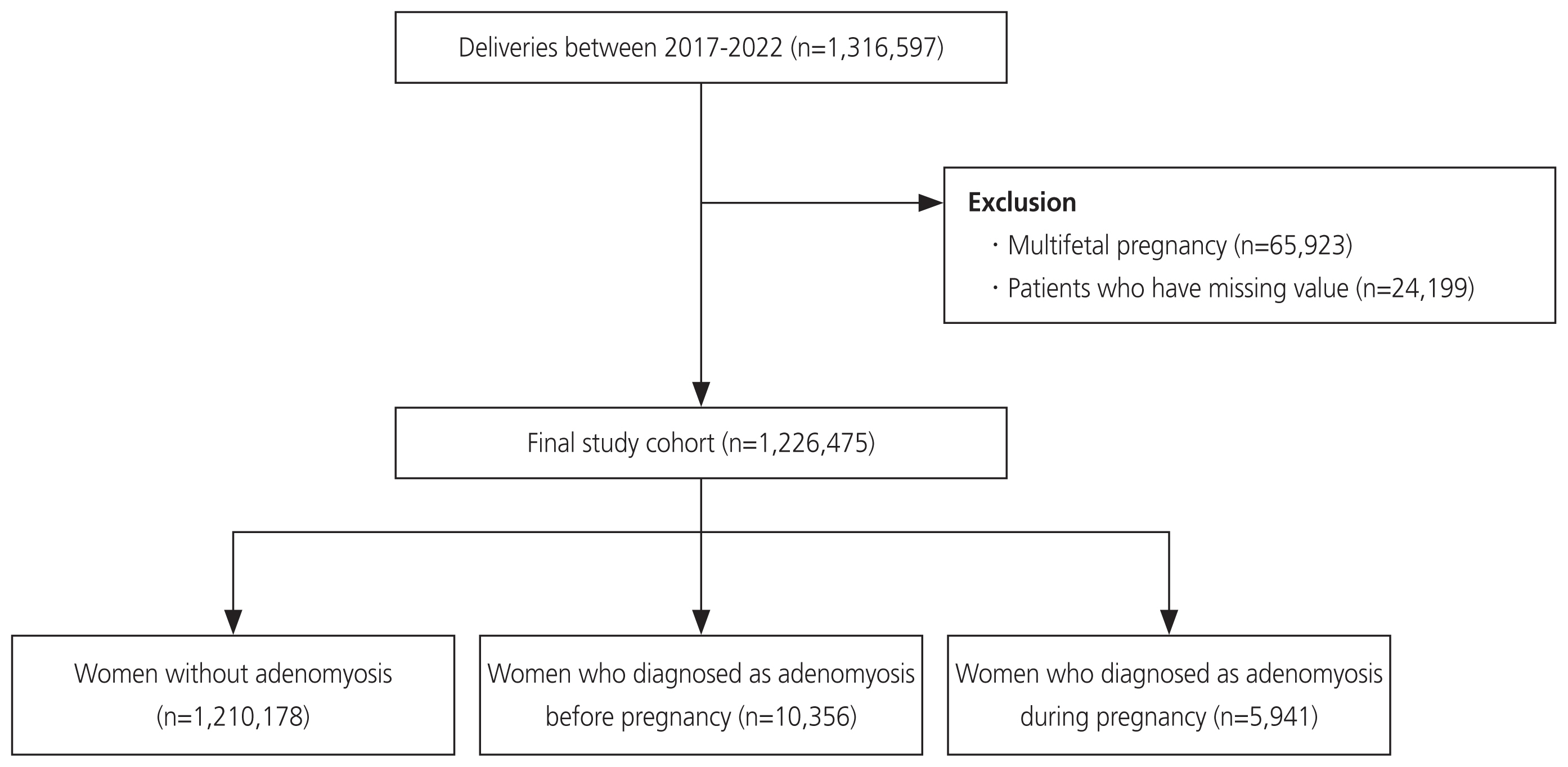
Flowchart of the study population.
3. Pregnancy and neonatal outcomes
Maternal health conditions were ascertained by querying the International Classification of Diseases 10th Revision (ICD-10) diagnosis codes. A diagnosis of maternal adenomyosis, both before and after pregnancy, was established when patients had been diagnosed with adenomyosis (ICD-10 code N80). Adenomyosis during pregnancy was confirmed through the identification of an ICD-10 code for the time during pregnancy, as there were no pre-pregnancy ICD-10 codes indicating its presence. Data on pregnancy outcomes were extracted using ICD-10 codes, which included information on the mode of delivery, underlying diseases, hypertensive disorder during pregnancy (HDP), gestational diabetes mellitus (GDM), postpartum hemorrhage (PPH), placental abruption, and placenta previa. Data on neonatal outcomes, including preterm birth and birth weight were extracted from the NHSP-IC database. Preterm birth was defined as having a gestational age <37 weeks, small for gestational age (SGA) was defined as a birthweight below the 10th percentile for the gestational age, and large for gestational age (LGA) was defined as a birthweight over the 90th percentile for the gestational age.
4. Statistical analysis
The continuous variables are presented as means and the standard deviation, and group comparisons were conducted using either Student’s t-test or the analysis of variance model for multiple groups. The categorical variables are presented as counts and percentages, and group comparisons were performed using the chi-square test. To assess the adverse pregnancy outcomes, a regression model was employed to calculate odds ratios (ORs) and their corresponding 95% confidence intervals. The statistical analyses were carried out using the SAS software version 9.4 for Windows (SAS Inc., Cary, NC, USA), and statistical significance was set at a P-value <0.05.
Results
1. Study population
Among the 1,316,597 women who delivered between 2017 and 2022, after excluding multiple pregnancies and missing data, a total of 1,226,475 women were included in the final analysis. Of these, 1,210,178 women had no diagnosis of adenomyosis (group 1), while 10,356 women were diagnosed with adenomyosis before pregnancy (group 2), and 5,941 women were diagnosed with adenomyosis during pregnancy (group 3) (Fig. 1). In Supplementary Table 1, the number of cases diagnosed with adenomyosis is presented annually.
Table 1 shows the baseline characteristics of the study population. The pregnant women with adenomyosis were older and more likely to be nulliparity than the pregnant women with no diagnosis of adenomyosis. The women in groups 2 and 3 had higher prevalence of hypertension before pregnancy and a history of overt diabetes compared with those in group 1, but there was no statistically significant difference between group 2 and group 3 (hypertension before pregnancy, 0.89% in group 1; 1.89% in group 2; and 1.57% in group 3, P<0.0001; overt diabetes mellitus, 1.76% in group 1; 3.34% in group 2; 2.54% in group 3, P<0.0001).

Baseline characteristics of the study population
2. Pregnancy and neonatal outcomes
Table 2 presents the pregnancy and neonatal outcomes of the three groups. The pregnant women with adenomyosis had an increased risk of adverse pregnancy outcomes including HDP, GDM, cesarean section, PPH, placenta previa, placental abruption, preterm delivery, SGA, and LGA compared with those in group 1. For groups 2 and 3, the occurrence of GDM and preterm labor was higher in group 2 compared with group 3.
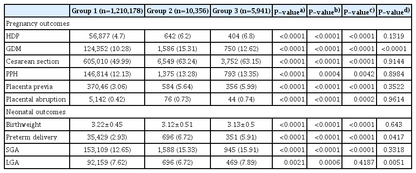
Pregnancy outcomes and neonatal outcomes according to the timing of adenomyosis diagnosis
3. Risk of adverse pregnancy outcomes
Table 3 summarizes the ORs of the presence of adenomyosis before pregnancy for adverse pregnancy outcomes such as HDP, GDM, cesarean section, PPH, placenta previa, placental abruption, preterm delivery, SGA, and LGA compared with those with no adenomyosis or adenomyosis diagnosed during pregnancy after adjustment for confounding variables.

Multivariate analyses for pregnancy and neonatal outcomes
In group 3, HDP, the risks of cesarean section, PPH, placenta previa, placental abruption, and SGA were the highest. HDP and the risk of cesarean section exhibited a statistically significant difference between groups 2 and 3. In group 2, the risks of GDM and preterm delivery were the highest. Interestingly, for GDM, the risk was found to decrease in group 3 compared to group 2, and for preterm delivery, there was no statistically significant difference between the two groups.
Discussion
The main findings of this study were 1) women with a diagnosis of adenomyosis had significantly higher risk of adverse pregnancy outcomes; 2) HPD, the risks of cesarean section, PPH, placenta previa, placental abruption, and SGA were all highest in cases with diagnosed adenomyosis during pregnancy, and HDP and cesarean section exhibited a statistically significant difference between groups 2 and 3; and 3) conversely, the risks of GDM and preterm delivery were highest risk in the group diagnosed with adenomyosis before pregnancy. However, only GDM exhibited a statistically significant difference between groups 2 and 3.
Previous research has shown an increased risk of adverse pregnancy outcomes in women with adenomyosis. These outcomes include a higher likelihood of preterm delivery, fetal malpresentation, postpartum hemorrhage, preeclampsia, low birth weight, and having a small-for-gestational-age newborn [3,5,7]. Additionally, women with adenomyosis have been found to have an elevated risk of miscarriage and a reduced chance of achieving a live birth [8,9]. While the severity and timing of adenomyosis diagnosis can influence the extent of these adverse outcomes, the collective evidence underscores the importance of close monitoring and tailored care for pregnant individuals with adenomyosis to optimize pregnancy outcomes.
The impact of adenomyosis on pregnancy outcomes can vary depending on the region and extent of adenomyotic involvement. Women with diffuse or extensive forms of adenomyosis have been reported to have a higher risk of adverse pregnancy outcomes, including preterm delivery, postpartum hemorrhage, fetal malpresentation, and preeclampsia [6,10]. This suggests that widespread distribution of adenomyotic lesions within the uterine wall may lead to greater uterine dysfunction and complications during pregnancy [11]. However, it is important to note that the specific regional characteristics and extent of adenomyosis can influence the extent of its impact, with diffuse forms generally associated with more pronounced adverse outcomes.
In the current study, the individuals diagnosed with adenomyosis before pregnancy had an increased risk of some adverse pregnancy outcomes. Typically, those diagnosed with adenomyosis before pregnancy might have had more severe symptoms or a broader disease extent, making it easier to diagnose through methods such as ultrasound or magnetic resonance imaging. The pathogenic mechanisms underlying the impact of adenomyosis on the course of pregnancy are multifaceted. Adenomyosis can disrupt the uterine junctional zone (JZ), thereby affecting uterine peristalsis during the luteal phase, which is crucial for successful implantation [12]. This abnormal uterine contractility has been associated with conditions such as placenta previa and accreta, as well as uterine hyperstimulation, atony, placental retention, and postpartum hemorrhage [4,13]. Additionally, adenomyosis can increase intrauterine oxidative stress, thereby leading to maternal endothelial dysfunction, which underlies abnormal placentation. Such oxidative stress can result in hyperplastic changes in the spiral arteries, thereby increasing flow impedance in the uterine arteries and contributing to placentation defects [14]. Furthermore, the inflammatory environment associated with adenomyosis can alter myometrial decidualization and disrupt trophoblastic JZ invasion during pregnancy [15]. These complex pathogenic mechanisms shed light on how adenomyosis can adversely affect pregnancy outcomes, including preeclampsia, preterm delivery, fetal malpresentation, postpartum hemorrhage, low birth weight, and small-for-gestational-age infants. Understanding these mechanisms is key to improving the care and management of pregnant individuals with adenomyosis [16].
This study, using a large-scale national dataset, investigated pregnancy and neonatal outcomes based on the timing of adenomyosis diagnosis, providing additional evidence regarding the existing research on disease severity and extent. However, as a retrospective study, this research has limitations, and there may be constraints associated with defining the disease using ICD codes. Additionally, due to the nature of the data, it was not possible to assess the impact of the mode of conception, which can influence pregnancy outcomes. Furthermore, we could not ascertain the severity of adenomyosis, as our analysis was based on diagnostic codes and the timing of diagnosis.
In conclusion, women with a diagnosis of adenomyosis had a significantly higher risk of adverse pregnancy outcomes. The timing of adenomyosis diagnosis had varying risk levels depending on the type of pregnancy and neonatal outcomes. Therefore, efforts for more careful monitoring and prevention of adverse outcomes may be necessary when such women become pregnant.
Supplementary Information
Notes
Conflicts of interest
None to declare.
Ethical approval
The study’s protocol received approval from the Institutional Review Board of the Korea University Guro Hospital (2023GR0532).
Patient consent
Patient consent was waived by the IRB due to the retrospective nature of the study.
Funding information
Not applicable.