


Introduction
Laparoscopic myomectomy is more useful in shortening the length of hospital stay and reducing postoperative pain compared to laparotomy. It is also advantageous over laparotomy in that it produces less pelvic adhesion, which is remarkable discovery for patients desiring fertility. However, there are an increasing number of reports about uterine rupture or scar dehiscence during subsequent pregnancy in patients with a history of taking laparoscopic myomectomy. In this report, we present two cases of uterine scar dehiscence during pregnancy after laparoscopic myomectomy.
Case report
1. Case 1
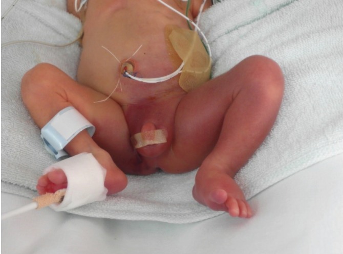
A 35-year-old nulligravida was admitted with a complaint of abdominal discomfort at 26 weeks of gestation. She had a 3-month history of laparoscopic myomectomy before conception. On admission, she had normal vital signs and laboratory findings. Non-stress test showed moderate fetal heart rate variability with irregular uterine contractions. On sonographic evaluation, however, we found that there was a 2.9-cm-sized defect on the left uterine wall. The fetal membrane was protruded from the defect but the placenta, umbilical cord and the fetus seemed intact. We decided to prolong the pregnancy and thus started tocolytic treatment. At 28 weeks of gestation, follow up ultrasound showed that the fetus had a leg stretched through the defect. Through emergency cesarean section, a 1,200-g male newborn was delivered and we found a 4-cm-sized uterine defect through which fetal leg protruded. We repaired the defect using a 2-layer sutures. Initially, the baby had a bruise and an edema on the left leg(Fig. 1) but eventually recovered during hospitalization. The patient underwent uneventful postoperative course and was discharged.
2. Case 2
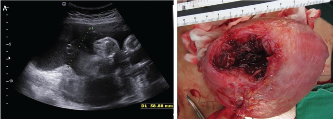
A 40-year-old primigravida visited us, complaining abdominal pain at 31 weeks of gestation. She had a 2-year-history of taking laparoscopic myomectomy. At 10 months thereafter, one year before this pregnancy, she had delivered the first baby by elective cesarean section. Intraoperative findings showed the thinning of the right fundal uterine wall where the myoma had been removed previously. On admission, while other test results were within normal range, ultrasound showed the myomectomy scar dehiscence (Fig. 2A). Since there were no findings suggestive of fetal distress, we decided to carry out the conservative management initially, but the pain aggravated despite a few hours use of tocolytics. On follow-up ultrasound, fetal leg was protruded through the defect as case 1. Through an emergency cesarean section, she delivered a 1,460-g baby with Apgar scores of 8 and 9 at 1 and 5 minutes, respectively. Intraoperatively, there was a 5 cm sized uterine defect (Fig. 2B) with protrusion of fetal leg, which was repaired using a 2-layer suture technique. Previous cesarean section scar was intact. The newborn did not show any leg abnormalities but had respiratory distress due to prematurity and the mother had uneventful postoperative course.
Discussion
Uterine scar dehiscence, defined as an asymptomatic thinning or separation of a prior uterine scar, is a rare complication that may occur following laparoscopic myomectomy [1]. Since patient with a history of uterine manipulation such as curettage to myomectomy or cesarean section are vulnerable to uterine wall weakness through scar site, they are at increased risk of developing uterine scar dehiscence [2]. According to a review of literatures, the risk of uterine rupture during subsequent pregnancy and delivery was up to 5.3% after laparotomy myomectomy [3,4] and 0.1% to 1% after laparoscopic myomectomy [4,5] respectively.
Major risk factors of developing scar dehiscence following laparoscopic myomectomy include a use of electrocautery and a lack of proper repair. Many studies have concluded that an excessive use of electrocautery may cause uterine dehiscence even with myomas confined to subserosal layers [1,2,6]. Suture technique is found to be more important factor for proper wound closure than the number of suture layers. Recent data has suggested that a 2-layer suture technique is not superior to a 1-layer one in correcting the separation of the uterine wall layers [7]. In case 2, the patient experienced uterine scar dehiscence in second pregnancy but her first was uneventful. We suspect the reason for uterine vulnerability that led to dehiscence is increased parity. In fact, numerous studies have suggested that high parity might be a risk factor for uterine rupture [8,9].
To date, several cases regarding a conservative management of uterine dehiscence during the second trimester have been reported. In some of these cases, tocolytics were given in order to seize uterine contraction [1] or to relieve abdominal pain [10,11]. In our two cases, we used tocolytics to prolong the pregnancy by alleviating the patients' symptoms because of fetal prematurity. Both patients were stable at the time of decision for conservation, and there were no identifiable contraindications of tocolytic use. In the first case, we were able to prolong the pregnancy for two weeks, but in the second case, as well as in other case reports, tocolytics failed to prolong gestation [1,10,11]. As there are no clinical recommendations for the use of tocolytics in conservative management of uterine dehiscence, each patients should be managed on a case by case basis.
There is still a controversy as to whether clinicians should prolong the pregnancy after detection of uterine scar dehiscence in preterm gestations [12]. Then, clinicians should also consider factors such as symptoms and signs of maternal or fetal distress, the location and the size of the uterine dehiscence and the distance between placenta and the dehiscence. In our cases, the patients delivered preterm babies through an emergency cesarean section because of the herniation of fetal parts through the dehiscence. Finally, clinicians should also consider other risk factors that might cause obstetric emergency, including early rupture of membrane and cord complications at the herniation site.


")


