Obstet Gynecol Sci Search

CLOSE


Obstet Gynecol Sci > Volume 65(4); 2022 > Article
Park, Kim, Kim, Kim, and Yim: Safety and feasibility of laterally extended endopelvic resection for sarcoma in the female genital tract: a prospective cohort study

Abstract

Objective

This study aims to evaluate the safety and feasibility of laterally extended endopelvic resection (LEER) for sarcoma in the female genital tract.

Methods

We prospectively recruited gynecologic cancer patients with sarcoma arising from female genital tract who underwent LEER at Seoul National University Hospital from December 2016 to March 2021. Clinicopathologic characteristics, surgical outcomes including postoperative complications and pain control, and survival outcomes of the patients were investigated.

Results

A total of nine patients were registered for this study. The median age was 56 years. Carcinosarcoma (n=2, 22%), leiomyosarcoma (n=2, 22%), and undifferentiated uterine sarcoma (n=2, 22%) were common histology types. Complete resection was achieved in 88.9%. The most common location of pelvic sidewall tumors was infra-iliac acetabulum (66.7%). The pathologic outcome showed a median tumor size of 9.0 cm and internal iliac vessel resection with pelvic sidewall muscle was performed in all patients. The median estimated blood loss was 1,600 mL (range, 300-22,300), and the patients were postoperatively admitted to the intensive care unit for median 1 day (range, 0-8). Complete response was observed in 44.4% (4/9) in radiologic studies after LEER, and median progression-free survival, treatment-related survival, and overall survival were 3.3, 19.6, and 98.9 months, respectively.

Conclusion

LEER was feasible and safe in treating recurrent sarcoma presenting pelvic sidewall invasion with acceptable survival outcomes and manageable postoperative complications.

Introduction

Sarcomas of the female genital tract are rare mesenchymal neoplasms, accounting for 1-3% of female genital tract malignancies [1]. Among them, uterine sarcomas contribute to 3-7% of uterine malignancies [2,3] and ovarian sarcomas contribute to 1% of ovarian malignancies [1,4,5]. Among gynecological sarcomas, uterine sarcoma is the most common (83%), followed by ovarian sarcoma (8%) [6]. Recent updates on female genital tract pathology have classified uterine sarcomas as mesenchymal tumors specific to the uterus, and the sub-classifications are as follows: leiomyosarcoma, endometrial stromal sarcoma (low grade), endometrial stromal sarcoma (high grade), and undifferentiated sarcoma [7]. Before the year 2014, carcinosarcoma had been classified as uterine sarcoma. However, in the 2014 World Health Organization classification it was reclassified as uterine carcinoma based on the tumor biology [8]. Nevertheless, due to its aggressive behavior, carcinosarcoma is still classified and analyzed together with sarcoma in several works of literature [3]. Five-year relative survival rate of uterine sarcoma is 40-50% [2], and poor survival rate of 37.33% and 12.25% have been reported in regional and distant stages, respectively [9]. Despite the poor prognosis, the role of surgery in the initial treatment stage of sarcoma patients is crucial in terms of diagnosis and survival. Preoperative diagnosis of sarcoma originating from female genital tract using imaging or cytology is difficult, and most gynecologic sarcomas can be diagnosed accurately only after surgery followed by careful pathologic examinations [10,11]. So far, the International Federation of Gynecology and Obstetrics (FIGO) staging and the absence of residual tumor after surgery are known prognostic factors for uterine sarcoma [10,12,13]. Furthermore, the role of adjuvant treatment, including radiotherapy or chemotherapy after surgery, is controversial [9,14,15]. Moreover, complete resection during secondary cytoreductive surgery has been shown to be effective for recurrent gynecologic sarcomas [16]. Therefore, complete cytoreduction is required for gynecologic sarcomas in both primary and recurrent settings.
Laterally extended endopelvic resection (LEER) was introduced in 1999 to treat recurrent cervical cancer involving the pelvic sidewall [17]. The LEER procedure enabled gynecologic surgeons to remove tumors at the pelvic sidewall, achieving R0 resection, which could not be achieved with pelvic exenteration alone. In previous studies, only heterogeneous data containing a small number of sarcomas were presented [18,19]. Therefore, this study aims to conduct an in-depth case review of gynecologic sarcoma who underwent LEER at Seoul National University Hospital.

Materials and methods

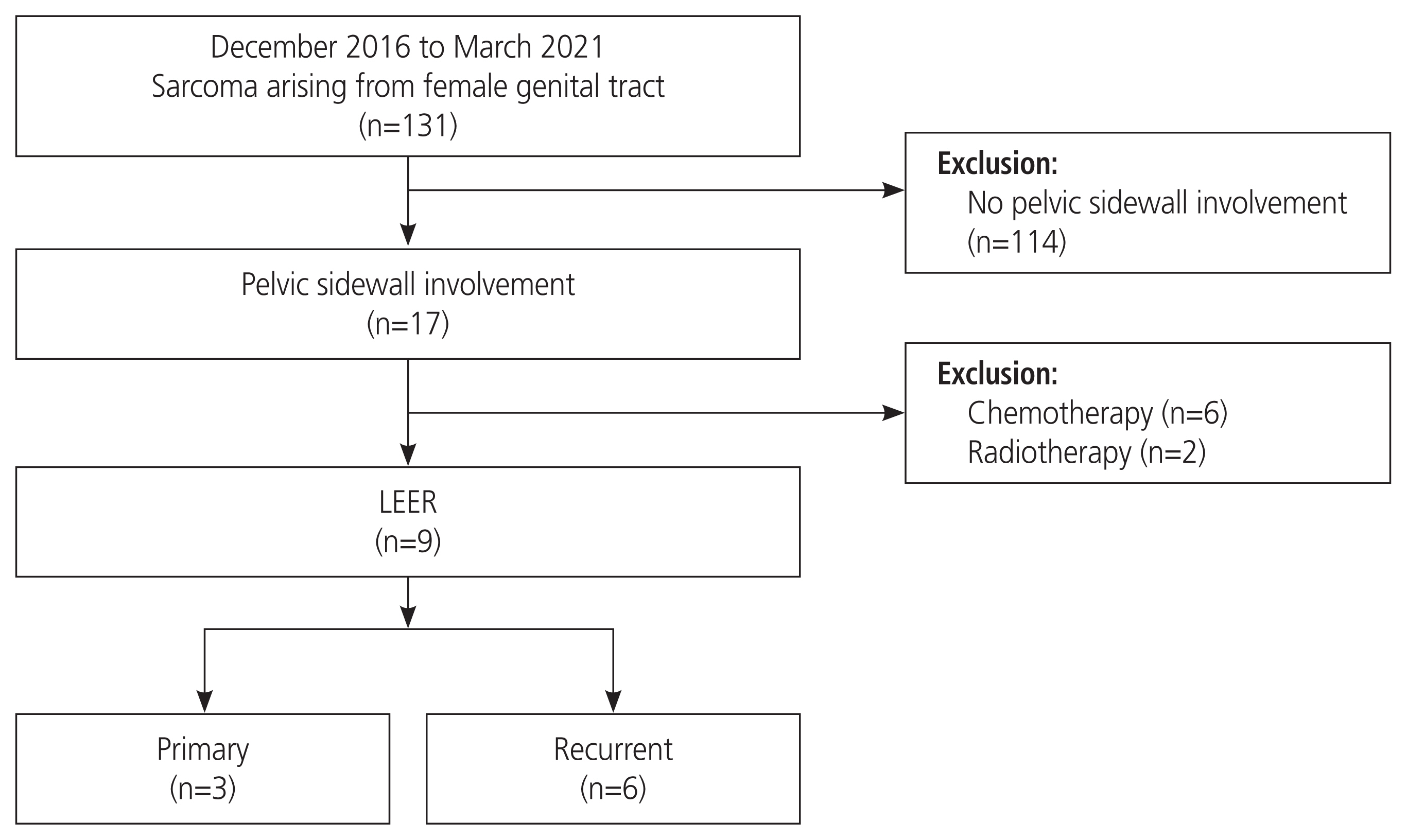
We prospectively collected data of patients with pelvic wall sarcoma who underwent LEER at Seoul National University Hospital between December 2016 and March 2021 (Fig. 1). This study was registered in the public registry (ClinicalTrials. gov identifier: NCT02986568). We included patients with the following features: 1) age of 20 years or older, 2) primary or recurrent pelvic wall sarcoma, 3) pelvic wall sarcoma without the involvement of the ipsilateral sciatic foramen, and 4) pelvic wall sarcoma expected to be cured by LEER. Among them, we excluded patients if they had bilateral pelvic wall sarcoma or any other available treatment options except LEER. We collected clinicopathologic data, including age, comorbidity, histologic type, disease status at the time of surgery, initial FIGO stage or American Joint Committee of Cancer soft tissue sarcoma staging system [20], preoperative lesion size of pelvic sidewall tumors, disease extent assessed by The TNM classification of malignant tumors (TNM) stage on radiologic imaging studies [21], topographic location of pelvic sidewall tumors, types of previous treatment, lines of prior chemotherapy, prior chemotherapy regimen and cycles, prior targeted-agent therapy, treatment-free interval before LEER, and median duration of follow-up. We also collected perioperative data associated with pathology and surgical outcomes, including surgery, pathologic tumor size, tumor grade, residual tumor, operation time, estimated blood loss (EBL), transfusion, intensive care unit (ICU) admission days, postoperative complications according to the Memorial Sloan Kettering Cancer Center (MSKCC) criteria, pre- and postoperative pain intensity assessed by numerical rating scale (NRS) and morphine milligram equivalents (MME), and types of postoperative adjuvant treatment provided. Data on survival outcomes were also collected, including treatment response at postoperative 3 months by radiologic examination, including computed tomography (CT) or magnetic resonance imaging, progression free survival (PFS), treatment-related survival (TRS), and overall survival (OS) from the day of diagnosis of the disease.

1. Procedures and treatment

LEER was performed as previously described [22-24]. The main procedures are as follows: 1) midline incision; 2) bowel mobilization; 3) dissection of both ureters; 4) skeletonization of the mesosigmoid colon and mesorectum; 5) en bloc resection of pelvic sidewall tumors with ligation of the internal iliac artery and vein below the bifurcation level of the common iliac artery; 6) pelvic muscle (obturator internus muscle, coccygeus muscle, pubococcygeus muscle, and iliococcygeus muscle) resection depending on tumor involvement and topography; 7) resection of urethra, lower vagina, and anus through vulva incision; and 8) permanent colostomy or ileal conduit. Organ preservation was considered if the resection margin was negative in the frozen section. Complete resection (R0) was defined as the absence of tumor in the lateral margins of all the resected tissues on the pathologic report. Postoperative complications were assessed by the MSKCC criteria [25]. Then, we performed adjuvant radiotherapy according to the radiation oncologist’s suggestion and administered chemotherapy to patients with distant metastasis.

2. Outcomes

The primary outcomes were tumor response at postoperative 3 months after LEER, PFS after LEER, TRS after LEER, and OS. Tumor response at postoperative 3 months after LEER was defined as disease status on abdominal or pelvic CT at 3 months after LEER. In this study, PFS after LEER was defined as the duration from operation to the day of first encountering disease progression, as confirmed by radiologic imaging studies. TRS after LEER was defined as the duration from the day of operation to the day of the last follow-up or death. OS was defined as the duration from the day of diagnosis of the disease to the day of the last follow-up or the day of death of the patient. We assumed all patients died a month after the last follow-up if they had a hopeless discharge with progressive disease. The secondary outcomes were perioperative characteristics: residual tumor, operation time, EBL, transfusion, postoperative ICU admission days, postoperative complications, and pre- and postoperative pain intensity. We assessed tumor response using the revised Response Evaluation Criteria in Solid Tumors version 1.1 [26]. Pelvic pain intensity was evaluated using both NRS and MME dose [27].

3. Statistical analysis

We analyzed patients’ clinicopathologic characteristics and surgical outcomes via descriptive statistics. Survival data, including PFS, TRS, and OS, were calculated by Kaplan-Meier survival analysis. SPSS software version 26.0 (SPSS Inc., Chicago, IL, USA) was used for statistical analysis in this study.

Results

1. Study population

In total, nine patients were included in this study. Table 1 shows patient characteristics. The median age of the study participants was 56 years (range, 22-65). Carcinosarcoma (n=2, 22%), leiomyosarcoma (n=2, 22%), and undifferentiated uterine sarcoma (n=2, 22%) were the common histologic types. Before surgery, the largest median radiologic tumor size was 10 cm (range, 2-17.5). Most patients with recurrent disease had received adjuvant chemotherapy or radiotherapy along with or without surgery. The median lines of prior chemotherapy before LEER were two (range, 0-5). The common chemotherapy regimen before LEER was an ifosfamide-combined regimen (n=5, 62.5%), such as doxorubicin-ifosfamide, paclitaxel-ifosfamide, and ifosfamidecisplatin. One patient (11.1%) had received targeted agent therapy (pazopanib). The median treatment free interval before LEER was 3.9 months (range, 1.1-38.2). The most common location of the pelvic sidewall tumor was the infra-iliac acetabulum (66.7%). Distant metastasis was observed in one patient (11.1%). The median duration of follow-up was 54.7 months (range, 11.4-130.4).

2. Surgical outcomes and perioperative complications

Surgical outcomes are shown in Table 2. The median tumor size in the pathologic report was 9.0 cm (range, 1.8-19.0). Complete resection was achieved in seven patients (88.9%). Organ preservation was achieved in eight patients (77.8%). Most patients showed high-grade pathology (n=8, 88.9%). The median operation time was 300 minutes (range, 135-1,320 minutes) with a median EBL of 1,600 mL (range, 300-22,300). Patients who underwent LEER were postoperatively admitted to the ICU for a median of 1 day (range, 0-8). The most common postoperative complication was peripheral neuropathy of grade 1 or 2 (44.4%), categorized as a nervous system complication according to the MSKCC criteria. Grade 1 or 2 genitourinary complications and infections were the next common postoperative complications (22.2%). Preoperative median pain intensity assessed by NRS was 4 (range, 0-7), and postoperative NRS was 2 (range, 1-3). The median preoperative MME was 0 mg/day (range, 0-105) and the postoperative MME was 0 mg/day (range, 0-15). Chemotherapy was the most favored postoperative adjuvant treatment (44.4%).

3. Treatment response and survival

Treatment response and survival outcomes after LEER are shown in Table 3. Treatment response at postoperative three months after LEER assessed by imaging studies showed a complete response (CR) in four (44.4%) patients, progressive disease (PD) in four patients (44.4%) and was not assessable in one patient. The median PFS after LEER was 3.3 months (95% confidence interval [CI], 2.4-4.1 months). The median OS was 98.9 months (95% CI could not be calculated due to censoring). The median TRS was 19.6 months (95% CI, 0-50.8).
All nine patients were individually investigated to identify potential risk factors for poor survival associated with LEER, as presented in Table 3. Two patients showed no progression until their last follow-up, and two achieved CR (22.2%). Their histologic types were low-grade endometrial stromal sarcoma (LGESS) and synovial sarcoma. Notably, the patient with LGESS did not achieve complete resection due to positive resection margin but showed CR at follow-up with no recurrence yet. Synovial sarcoma patient was the only one in this study who achieved CR just after surgery. Both patients were at an advanced stage. However, they were much younger than the patients with progressive disease, and aged 25 and 22 years, respectively. Although not described in the table, three patients (33.3%) were treated for pelvic sidewall recurrence with a targeted agent or chemotherapy before LEER. All these patients showed PD as their best response.

Discussion

This study shows the clinical outcome of LEER in female genital tract sarcoma patients with pelvic sidewall invasion and various histologic types. A high R0 rate of 88.9% was achieved without severe complications. While the involvement of major vessels and the pelvic sidewall often impedes the decision of surgical resection, the LEER procedure enables gynecologic surgeons to resect pelvic sidewall tumors to achieve a higher rate of R0 resection [18,27]. Surgical resection of localized recurrent disease has significantly improved the local control of soft tissue sarcoma [28,29]. However, till date, there is insufficient evidence regarding the role and safety of aggressive surgery in the localized recurrences of female genital tract sarcomas. Moreover, previous reports on LEER mainly focused on carcinoma patients, and in a study with sarcoma patients, the surgical and survival outcomes were not specifically reported [18].
The importance and indication of LEER surgery in sarcoma is described in the following text. First, R0 en bloc resection is essential for improving prognosis in sarcoma [30,31]. In particular, it is known that sarcomas grow in anatomic compartments and do not easily invade local anatomical boundaries [32,33]. The biological features of sarcomas enable R0 resection, coincidentally in accordance with Michel Höckel’s ontogenetic field theory [34]. In our prospective cohort study, eight patients achieved microscopic R0 in all surgical specimens, however, one patient (patient 1) with LGESS showed a positive resection margin. However, patient 1 was successfully treated with adjuvant hormonal therapy with letrozole and survived for 68.7 months without recurrence after LEER.
Second, the histologic type should be considered before surgery. We included various histologic types of recurrent disease, including leiomyosarcoma, undifferentiated uterine sarcoma, carcinosarcoma, and adenosarcoma. Leiomyosarcoma is the most common uterine sarcoma; and the beneficial effect of secondary surgical resection of uterine leiomyosarcoma has been shown in comparative retrospective cohort studies [16,35]. In our study, two patients with leiomyosarcoma were included: one had an OS of 65 months, and the other showed loss of follow-up but no evidence of recurrence. However, undifferentiated uterine sarcoma is a rare histologic type with an aggressive nature compared to other histologies [36]. Two patients (patients 7 and 8) with undifferentiated uterine sarcoma were included in our study, and they showed rapid progression within 2 months and 3.1 months, respectively, after LEER. Recurrent uterine carcinosarcoma also presents a poor prognosis. Systemic treatment showed a median PFS of 1.8 months [37], while patients 4 and 6 showed 3.3 and 2.6 months of PFS after LEER. Therefore, the decision for surgical excision in recurrent sarcoma should be appropriately made according to the surgeon’s judgment in consideration of the histologic type and the extent of surgery.
Third, tumors in the pelvic sidewall cause neuropathic pain due to sciatic nerve compression or irritation [27,38]. In this study, we found no statistically significant reduction in pain due to the small sample size. However, in our previous study, we reported that LEER significantly reduced pelvic sciatic pain and morphine requirements in patients with recurrent cervical cancer compared to chemotherapy [27]. However, before surgery, 55.9% of patients complained of moderate to severe pain with an NRS score ≥4; after surgery, the pain intensity decreased to an NRS score ≤3. Moreover, there were no grade 3/4 complications requiring invasive intervention in our cohort in terms of postoperative complications. A previous study had reported 22-28% of grade 4 complications after LEER [39,40].
Based on our clinical experience with a prospective cohort of patients with sarcoma and recurrent cervical cancer [24], R0 resection can be achieved after careful review of physical and radiologic findings. As described by Höckel [17], LEER is contraindicated in pelvic sidewall tumors involving the external iliac vessels. In addition, patients showing rectovaginal or vesicovaginal fistula may be candidates for LEER as palliative intent with or without distant metastasis [24]. Finally, uncontrolled pain requiring excessive opioids may be an indication for LEER based on our recurrent cervical cancer cohort study [24].
The strength of our study is the prospective nature of the studied cohort. Our study is of substantial value because gynecologic sarcomas and pelvic sidewall recurrences are rare. The limitations of our study are as follows: first, the sample size of the study was small, histologic types were heterogeneous, and primary and recurrent diseases were analyzed together. It was difficult to evaluate the potential prognostic factors of sarcoma treatment involving the pelvic sidewall. Second, it was challenging to compare the efficacy in terms of oncologic outcomes and treatment related complications due to the lack of a comparison cohort. Therefore, a multicenter, large-scale, cohort-based study of gynecologic sarcoma is warranted.
To date, there is no literature on the surgical treatment of sarcoma invading the pelvic sidewall, and this is the first prospective cohort study on LEER. In conclusion, LEER may be a feasible treatment option for gynecologic sarcoma and R0 resection can be attempted in tumors with pelvic sidewall invasion with acceptable oncologic and safety outcomes.

Acknowledgments

We thank the FUSION study group at Seoul National University Hospital for participating in the collaborative work on LEER. We deeply appreciate Dreampac Corp. (Wonju, Korea) and Precision Medicine for Peritoneal Metastasis Corp. (Wonju, Korea) for their support.

Notes

Conflict of interest

No potential conflict of interest relevant to this article was reported.

Jae-Weon Kim has been an Editorial Board of Obstetrics & Gynecology Science; however, he was not involved in the peer reviewer selection, evaluation, or decision process of this article. Otherwise, no other potential conflicts of interest relevant to this article were reported.

Ethical approval

Seoul National University Institutional Review Board has approved this study (IRB No: 1506-113-682).

Patient consent

All patients signed the informed consent form.

Funding information

None.

Fig. 1
Patient flowchart. LEER, laterally extended endopelvic resection.
ogs-22071f1.jpg
Table 1
Patient characteristics
Characteristic Value (n=9)
Age (yr) 56 (22-65)
Comorbidity
 Yesa) 5 (55.5)
 None 4 (44.4)
Histology
 Carcinosarcoma 2 (22.2)
 Leiomyosarcoma 2 (22.2)
 Undifferentiated uterine sarcoma 2 (22.2)
 Low-grade endometrial stromal sarcoma 1 (11.1)
 Mullerian adenosarcoma 1 (11.1)
 Synovial sarcoma 1 (11.1)
Disease status at the time of LEER
 Primary disease 3 (33.3)
 Recurrent disease 6 (66.7)
Initial FIGO stage
 I 4 (44.4)
 II 1 (11.1)
 III 2 (22.2)b)
 IV 2 (22.2)
Preoperative radiologic TNM stage
 T staging
  T2b 5 (55.6)
  T4 4 (44.4)
 N staging
  N0 6 (66.7)
  N1 3 (33.3)
 M staging
  M0 7 (77.8)
  M1 2 (22.2)
Largest radiologic tumor size prior to LEER (cm) 10 (2-17.5)
Neoadjuvant chemotherapyc) 2 (22.2)c)
Types of prior treatmentd)
 Systemic therapy 5 (55.5)
 Radiotherapy 2 (22.2)
 Surgery+chemotherapy 4 (44.4)
 Surgery+radiotherapy 2 (22.2)
Lines of prior chemotherapy 2 (0-5)
Prior systemic treatment regimend),e)
 Ifosfamide-combined 4 (44.4)
 Doxorubicin only or combined 3 (33.3)
 Gemcitabine-docetaxel 1 (11.1)
 Paclitaxel-carboplatin 1 (11.1)
 Targeted therapy 1 (11.1)
Treatment-free interval before LEER (months) 3.9 (1.1-38.2)
Best response of last treatment before LEER
 Complete response 2 (22.2)
 Partial response 2 (22.2)
 Stable disease 1 (11.1)
 Progressive disease 3 (33.3)
 Not available 1 (11.1)
Pelvic sidewall tumor locationd)
 Infra-iliac acetabulum 6 (66.7)
 Infra-iliac ischiopubic 2 (22.2)
 Infra-iliac sacrococcygeal 4 (44.4)
Duration of follow-up (months) 52.7 (11.4-130.4)

Values are presented as median (range) or number (%).

LEER, laterally extended endopelvic resection; FIGO, International Federation of Gynecology and Obstetrics; TNM, The TNM classification of malignant tumors.

a) Comorbidities included hypertension (n=1, 11.1%), diabetes (n=1, 11.1%), dyslipidemia (n=2, 22.2%), thyroid disease (n=1, 11.1%), thromboembolic disease (n=1, 11.1%), and hepatitis (n=1, 11,1%), overlapping conditions included;

b) Stage of the patient with synovial sarcoma was classified according to American Joint Committee of Cancer staging system (8th edition);

c) Neoadjuvant treatment among primary cases;

d) Overlapping cases were included;

e) Cases with neoadjuvant chemotherapy were included.

Table 2
Clinicopathologic and treatment outcomes of LEER and adjuvant treatment
Characteristic Value (n=9)
Organ preservation
 No 2 (22.2)
 Rectum alone 2 (22.2)
 Bladder alone 0 (0.0)
 Rectum and bladder both 5 (55.6)
Surgical extent
 Hysterectomy 2 (22.2)
 BSO 2 (22.2)
 PLND 7 (77.8)
 PALND 7 (77.8)
 Cystectomy 4 (44.4)
 Vaginectomy 4 (44.4)
 Internal iliac vessel resection 9 (100.0)
 Pelvic sidewall muscle resection 8 (88.9)
  Obturator internus muscle 6 (66.7)
  Iliococcygeus muscle 3 (33.3)
  Pubococcygeus muscle 5 (55.6)
  Coccygeus muscle 3 (33.3)
 Ureter ligation and resection 8 (88.9)
 Vulvectomy (perineum) 4 (44.4)
 Bowel resection 5 (55.6)
 Ileal conduit 4 (44.4)
 Colostomy 2 (22.2)
 Othersa) 5 (55.5)
Pathologic tumor size (cm) 9.0 (1.8-19.0)
Pathologic extent
 Uterus 2 (22.2)
 Vagina 4 (44.4)
 Perineum 0 (0.0)
 Bladder and urethra 7 (77.8)
 Anus and rectum 5 (55.6)
 Pelvic sidewall muscle 7 (77.8)
 Internal iliac vessel 6 (66.7)
Tumor grade
 Low-grade 1 (11.1)
 High-grade 8 (88.9)
Residual tumor
 R0 8 (88.9)
 R1 1 (11.1)
Operation time (minutes) 300 (135-1,320)
Estimated blood loss (mL) 1,600 (300-22,300)
Transfusion
 RBC 3 (0-42)
 FFP 0 (0-34)
 PC 0 (0-24)
Postoperative ICU admission (days) 1 (0-8)
Postoperative complications (according to MSKCC grading system)
 Gastrointestinal system (ileus)
  Grade 1/2 2 (22.2)
  Grade 3/4 0 (0.0)
 Genitourinary system (urinary incontinence, voiding difficulty)
  Grade 1/2 2 (22.2)
  Grade 3/4 0 (0.0)
 Infection
  Grade 1/2 0 (0.0)
  Grade 3/4 3 (33.3)
 Nervous system
  Grade 1/2 4 (44.4)
  Grade 3/4 0 (0.0)
Pelvic pain severity
 Preoperative NRS 4 (0-7)
 Postoperative NRS 2 (1-3)
 Preoperative MME (mg/day) 0 (0-105)
 Postoperative MME (mg/day) 0 (0-15)
Postoperative adjuvant treatment
 No adjuvant treatment 3 (33.3)
 Concurrent chemoradiation followed by hormone therapy 1 (11.1)
 Chemotherapy 4 (44.4)
 Concurrent chemoradiation 1 (11.1)
Treatment response at postoperative 3 months
 Complete response 4 (44.4)
 Partial response 0 (0.0)
 Stable disease 0 (0.0)
 Progression or recurrence 4 (44.4)
 Not assessable 1 (11.1)
Recurrence 6 (66.7)
Death 5 (55.6)

Values are presented as median (range) or number (%).

LEER, laterally extended endopelvic resection; BSO, bilateral-salpingo-oophorectomy; PLND, pelvic lymph node dissection; PALND, para-aortic lymph node dissection; RBC, red blood cell; FFP, fresh frozen plasma; PC, platelet concentrate; ICU, intensive care unit; MSKCC, Memorial Sloan Kettering Cancer Center; NRS, numeric rating scale; MME, morphine milligram equivalents.

a) Included partial cystectomy (n=3, 33.3%), liver resection (n=1, 11.1%), and video-assisted thoracoscopic lung lobectomy (n=1, 11.1%).

Table 3
Individual treatment information and outcomes of patients
Patient No. Cancer type Age at LEER Histology Tumor grade Initial FIGO stage Radiologic TNM stage Primary or recurrent Preoperative treatment Postoperative adjuvant treatment TFI (months) Tumor sites on pelvic sidewall Residual tumor Disease status at the last follow-up Treatment responses at postoperative three months Recurrence Death PFS after LEER (months) TRS after LEER (months) OS after diagnosis (months)
No progression at last follow-up
 1 UC 25 Low-grade endometrial stromal sarcoma Low-grade IVA T4N1M0 Primary Chemotherapy (doxorubicin-ifosfamide), CCRT (cisplatin) CCRT (cisplatin), hormone therapy (letrozole) 0 Infra-iliac sacrococcygeal R1 (margin+) CR CR No No 68.7 68.7 69.2
 2 UC 22 Synovial sarcoma High-grade III (AJCC) T2bN0M0 Primary None Chemotherapy (ifosfamide-doxorubicin), CCRT (cisplatin) 0 Infra-iliac acetabular, infra-iliac sacrococcygeal R0 CR CR No No 9.9 9.9 10.3
Progression or not assessable at last follow-up
 3 OC 45 Mullerian adenosarcoma High-grade IIIC T4N0M0 Recurrent Chemotherapy (paclitaxel-carboplatin) Chemotherapy, targeted therapy (liposomal doxorubicin-carboplatin; topotecan-bevacizumab; gemcitabine), radiotherapy 11.7 Infra-iliac sacrococcygeal R0 PD CR Yes No 10.2 40.2 98.9
 4 UC 55 Carcinosarcoma High-grade IB T4N1M0 Recurrent Chemotherapy (paclitaxel-Carboplatin), radiotherapy Chemotherapy (5 FU-cisplatin; ifosfamide-cisplatin) 1.3 Infra-iliac acetabular R0 PD CR Yes Yes 3.3 7.0 20.6
 5 UC 60 Leiomyosarcoma High-grade IB T4N0M1 Recurrent Chemotherapy (gemcitabine-docetaxel) Targeted therapy (pazopanib), chemotherapy (doxorubicin-cisplatin; ifosfamide-cisplatin; eribulin) 38.2 Infra-iliac sacrococcygeal R0 PD PD Yes Yes 0.3 20.6 65.0
 6 UC 59 Carcinosarcoma High-grade IB T2bN0M1 Recurrent Chemotherapy (ifosfamide-paclitaxel), CCRT (cisplatin) Chemotherapy (doxorubicin-cisplatin, 5 FU-cisplatin) 6.5 Infra-iliac ischiopubic, infra- iliac acetabular (Lt), infra-iliac ischiopubic (Rt) R0 PD PD Yes Yes 2.6 19.6 42.2
 7 UC 56 Undifferentiated uterine sarcoma High-grade IVA T4N1M0 Recurrent CCRT (cisplatin) None 1.1 Infra-iliac ischiopubic (Lt), infra-iliac acetabular (Rt) R0 PD PD Yes Yes 2.0 3.4 10.0
 8 UC 59 Undifferentiated uterine sarcoma High-grade IIB T2bN0M0 Primary Chemotherapy (doxorubicin-cisplatin) Chemotherapy (doxorubicin-cisplatin; ifosfamide-cisplatin) 0 Infra-iliac acetabular R0 PD PD Yes Yes 3.1 6.0 9.6
 9 UC 65 Leiomyosarcoma High-grade IB T2bN0M0 Recurrent Chemotherapy (ifosfamide-cisplatin), targeted therapy (pazopanib), CCRT (cisplatin) Chemotherapy (gemcitabine-docetaxel) 1.2 Infra-iliac acetabular R0 NA NA NA No 2.2 2.2 82.9

LEER, laterally extended endopelvic resection; FIGO, International Federation of Gynecology and Obstetrics; TNM, the TNM classification of malignant tumors; TFI, treatment free survival; PFS, progression free survival; TRS, treatment-related survival; OS, overall survival; UC, uterine cancer; CCRT, concurrent chemoradiation; CR, complete response; AJCC, American Joint Committee of Cancer; OC, ovarian cancer; PD, progressive disease; FU, follow up; Lt, left; Rt, right; NA, not available.

References

1. Cramer DW, Cutler SJ. Incidence and histopathology of malignancies of the female genital organs in the United States. Am J Obstet Gynecol 1974;118:443-60.
crossref pmid
2. Boll D, Verhoeven RH, van der Aa MA, Pauwels P, Karim-Kos HE, Coebergh JW, et al. Incidence and survival trends of uncommon corpus uteri malignancies in the Netherlands, 1989-2008. Int J Gynecol Cancer 2012;22:599-606.
crossref pmid
3. Mbatani N, Olawaiye AB, Prat J. Uterine sarcomas. Int J Gynaecol Obstet 2018;143 (Suppl 2):51-8.
crossref pmid pdf
4. Bacalbasa N, Balescu I, Dima S, Popescu I. Ovarian sarcoma carries a poorer prognosis than ovarian epithelial cancer throughout all FIGO stages: a single-center case-control matched study. Anticancer Res 2014;34:7303-8.
pmid
5. Ha HI, Chang HK, Park SJ, Lim J, Won YJ, Lim MC. The incidence and survival of cervical, ovarian, and endometrial cancer in Korea, 1999-2017: Korea central cancer registry. Obstet Gynecol Sci 2021;64:444-53.
crossref pmid pmc pdf
6. Francis M, Dennis NL, Hirschowitz L, Grimer R, Poole J, Lawrence G, et al. Incidence and survival of gynecologic sarcomas in England. Int J Gynecol Cancer 2015;25:850-7.
crossref pmid
7. Board WHO Classification of Tumours Editorial Board. Female genital tumours. Lyon: International Agency for Research on Cancer; 2020.

8. Kurman RJ, Carcangiu ML, Herrington CS, Young RH. WHO classification of tumours of female reproductive organs. Lyon: International Agency for Research on Cancer; 2014.

9. Hosh M, Antar S, Nazzal A, Warda M, Gibreel A, Refky B. Uterine sarcoma: analysis of 13,089 cases based on surveillance, epidemiology, and end results database. Int J Gynecol Cancer 2016;26:1098-104.
crossref pmid
10. Sagae S, Yamashita K, Ishioka S, Nishioka Y, Terasawa K, Mori M, et al. Preoperative diagnosis and treatment results in 106 patients with uterine sarcoma in Hokkaido, Japan. Oncology 2004;67:33-9.
crossref pmid pdf
11. Cho HY, Kim K, Kim YB, No JH. Differential diagnosis between uterine sarcoma and leiomyoma using preoperative clinical characteristics. J Obstet Gynaecol Res 2016;42:313-8.
crossref pmid pdf
12. Yim GW, Nam EJ, Kim SW, Kim YT. FIGO staging for uterine sarcomas: can the revised 2008 staging system predict survival outcome better? Yonsei Med J 2014;55:563-9.
crossref pmid pmc
13. Potikul C, Tangjitgamol S, Khunnarong J, Srijaipracharoen S, Thavaramara T, Pataradool K. Uterine sarcoma: clinical presentation, treatment and survival outcomes in thailand. Asian Pac J Cancer Prev 2016;17:1759-67.
crossref pmid
14. Rizzo A, Nannini M, Astolfi A, Indio V, De Iaco P, Perrone AM, et al. Impact of chemotherapy in the adjuvant setting of early stage uterine leiomyosarcoma: a systematic review and updated meta-analysis. Cancers (Basel) 2020;12:1899.
crossref pmid pmc
15. Vaz J, Tian C, Richardson MT, Chan JK, Mysona D, Rao UN, et al. Impact of adjuvant treatment and prognostic factors in stage i uterine leiomyosarcoma patients treated in commission on cancer®-accredited facilities. Gynecol Oncol 2020;157:121-30.
crossref pmid
16. Cybulska P, Sioulas V, Orfanelli T, Zivanovic O, Mueller JJ, Broach VA, et al. Secondary surgical resection for patients with recurrent uterine leiomyosarcoma. Gynecol Oncol 2019;154:333-7.
crossref pmid pmc
17. Höckel M. Laterally extended endopelvic resection: surgical treatment of infrailiac pelvic wall recurrences of gynecologic malignancies. Am J Obstet Gynecol 1999;180:306-12.
crossref pmid
18. Höckel M. Long-term experience with (laterally) extended endopelvic resection (LEER) in relapsed pelvic malignancies. Curr Oncol Rep 2015;17:435.
pmid
19. Cowie P, Eastwood B, Smyth S, Soleymani Majd H. Atypical presentation of intravascular leiomyomatosis mimicking advanced uterine sarcoma: modified laterally extended endopelvic resection with preservation of pelvic neural structures. BMJ Case Rep 2021;14 :e244774.
crossref pmid pmc
20. Cates JMM. The AJCC 8th edition staging system for soft tissue sarcoma of the extremities or trunk: a cohort study of the SEER database. J Natl Compr Canc Netw 2018;16:144-52.
crossref pmid
21. Brierley J, Gospodarowicz MK, Wittekind C. TNM classification of malignant tumours. 8th ed. West Sussex: John Wiley & Sons, Inc; 2017.

22. Höckel M. Laterally extended endopelvic resection (LEER)--principles and practice. Gynecol Oncol 2008;111:S13-7.
crossref pmid
23. Park SJ, Kim HS. Laterally extended endopelvic resection with nephrectomy for vaginal cancer. Gynecol Oncol 2019;152:218-9.
crossref pmid
24. Park SJ, Mun J, Lee S, Luo Y, Chung HH, Kim JW, et al. Laterally extended endopelvic resection versus chemo or targeted therapy alone for pelvic sidewall recurrence of cervical cancer. Front Oncol 2021;11:683441.
crossref pmid pmc
25. Strong VE, Selby LV, Sovel M, Disa JJ, Hoskins W, Dematteo R, et al. Development and assessment of memorial sloan kettering cancer center’s surgical secondary events grading system. Ann Surg Oncol 2015;22:1061-7.
crossref pmid pmc pdf
26. Eisenhauer EA, Therasse P, Bogaerts J, Schwartz LH, Sargent D, Ford R, et al. New response evaluation criteria in solid tumours: revised RECIST guideline (version 1.1). Eur J Cancer 2009;45:228-47.
crossref pmid
27. Dowell D, Haegerich TM, Chou R. CDC guideline for prescribing opioids for chronic pain--united states, 2016. JAMA 2016;315:1624-45.
crossref pmid pmc
28. Gronchi A, Lo Vullo S, Fiore M, Mussi C, Stacchiotti S, Collini P, et al. Aggressive surgical policies in a retrospectively reviewed single-institution case series of retroperitoneal soft tissue sarcoma patients. J Clin Oncol 2009;27:24-30.
crossref pmid
29. Radaelli S, Fiore M, Colombo C, Ford S, Palassini E, Sanfilippo R, et al. Vascular resection en-bloc with tumor removal and graft reconstruction is safe and effective in soft tissue sarcoma (STS) of the extremities and retro-peritoneum. Surg Oncol 2016;25:125-31.
crossref pmid
30. Gronchi A, Miah AB, Dei Tos AP, Abecassis N, Bajpai J, Bauer S, et al. Soft tissue and visceral sarcomas: ESMO-EURACAN-GENTURIS clinical practice guidelines for diagnosis, treatment and follow-up . Ann Oncol 2021;32:1348-65.
pmid
31. Asencio Pascual JM, Fernandez Hernandez JA, Blanco Fernandez G, Muñoz Casares C, Álvarez Álvarez R, Fox Anzorena B, et al. Update in pelvic and retroperitoneal sarcoma management: the role of compartment surgery. Cir Esp (Engl Ed) 2019;97:480-8.
crossref pmid
32. Anderson MW, Temple HT, Dussault RG, Kaplan PA. Compartmental anatomy: relevance to staging and biopsy of musculoskeletal tumors. AJR Am J Roentgenol 1999;173:1663-71.
crossref pmid
33. Enneking WF, Spanier SS, Malawer MM. The effect of the anatomic setting on the results of surgical procedures for soft parts sarcoma of the thigh. Cancer 1981;47:1005-22.
crossref pmid
34. Höckel M, Wolf B, Schmidt K, Mende M, Aktas B, Kimmig R, et al. Surgical resection based on ontogenetic cancer field theory for cervical cancer: mature results from a single-centre, prospective, observational, cohort study. Lancet Oncol 2019;20:1316-26.
crossref pmid
35. Leitao MM, Brennan MF, Hensley M, Sonoda Y, Hummer A, Bhaskaran D, et al. Surgical resection of pulmonary and extrapulmonary recurrences of uterine leiomyosarcoma. Gynecol Oncol 2002;87:287-94.
crossref pmid
36. Ríos I, Rovirosa A, Morales J, Gonzalez-Farre B, Arenas M, Ordi J, et al. Undifferentiated uterine sarcoma: a rare, not well known and aggressive disease: report of 13 cases. Arch Gynecol Obstet 2014;290:993-7.
crossref pmid pdf
37. Matsuzaki S, Klar M, Matsuzaki S, Roman LD, Sood AK, Matsuo K. Uterine carcinosarcoma: contemporary clinical summary, molecular updates, and future research opportunity. Gynecol Oncol 2021;160:586-601.
crossref pmid
38. Kanao H, Aoki Y, Fusegi A, Takeshima N. Should indications for laterally extended endopelvic resection (LEER) exclude patients with sciatica? J Gynecol Oncol 2020;31:e63.
crossref pmid pmc pdf
39. Kanao H, Aoki Y, Omi M, Nomura H, Tanigawa T, Okamoto S, et al. Laparoscopic pelvic exenteration and laterally extended endopelvic resection for postradiation recurrent cervical carcinoma: technical feasibility and short-term oncologic outcome. Gynecol Oncol 2021;161:34-8.
crossref pmid
40. Vizzielli G, Naik R, Dostalek L, Bizzarri N, Kucukmetin A, Tinelli G, et al. Laterally extended pelvic resection for gynaecological malignancies: a multicentric experience with out-of-the-box surgery. Ann Surg Oncol 2019;26:523-30.
crossref pmid pdf
TOOLS
Share :
Facebook Twitter Linked In Google+ Line it
METRICS Graph View
  • 2 Web of Science
  • 2 Crossref
  • 2   Scopus
  • 2,063 View
  • 82 Download
Related articles in Obstet Gynecol Sci


ABOUT
ARTICLE & TOPICS
Article category

Browse all articles >

Topics

Browse all articles >

BROWSE ARTICLES
POLICY
FOR CONTRIBUTORS
Editorial Office
4th Floor, 36 Gangnam-daero 132-gil, Gangnam-gu, Seoul 06044, Korea.
Tel: +82-2-2266-7238    Fax: +82-2-3445-2440    E-mail: journal@ogscience.org                

Copyright © 2024 by Korean Society of Obstetrics and Gynecology.

Developed in M2PI

Close layer
prev next