


Introduction
Adnexal masses are sometimes visualized during pregnancy, with an occurrence rate of 0.01-15% [1-9]. Their diagnosis and treatment planning can be challenging. Most of these masses are benign [10], and they spontaneously regress in 70% of cases [4,11]. However, approximately 1-8% of such masses are diagnosed as malignant [12,13]. Early detection of adnexal malignancy is crucial because 67% of patients show progressive disease and poor prognosis because of late detection [14,15]. Furthermore, with timely treatment, the oncologic outcomes of pregnant women have been reported to be similar to those of non-pregnant women with adnexal malignancy [16]. Therefore, risk assessment in pregnant women is important to ensure early diagnosis and timely management.
Adnexal masses in pregnant women are most commonly detected during ultrasonography (US) examination since it is routinely performed during antenatal care. Several US features and scoring systems [17-22] have been reported to be useful in predicting malignancy. Although US has its own merits for safety with optimal sensitivity and specificity, there are several limitations, including inter-observer variability and the difficulty in assessing adnexal masses over 20 weeks of gestation due to the augmented uterine volume. In non-pregnant women, computed tomography (CT) can be used to compensate for the limitations of US. According to a recent meta-analysis, CT shows good sensitivity and specificity in differentiating benign and malignant adnexal masses (79% and 87%, respectively) [23]. However, CT is contraindicated during pregnancy because of radiation toxicity, which can induce fetal malformations. Fluorine-18 fluoro-deoxy-glucose positron emission tomography/CT (18F-FDG PET/CT) also cannot be used for the same reason. Magnetic resonance imaging (MRI) is an alternative to CT and 18F-FDG PET/CT. MRI has shown better performance than CT in differentiating benign and malignant adnexal masses, with sensitivity and specificity of 94% and 91%, respectively [23]. A larger scanning area, better definition of tissue planes, and more information on tissue composition on MRI are the advantages of the technique over US in distinguishing adnexal masses. However, the high cost, long examination time with uncomfortable fixed supine position, and use of contrast agents are drawbacks of MRI. For clinicians to adequately manage patients with adnexal masses during pregnancy, it is crucial to understand both the US and MRI findings to predict the risk of malignancy.
Therefore, we thoroughly reviewed and described the characteristic imaging features of benign and malignant adnexal masses detected in pregnant women using US and MRI.
Diagnostic evaluation on adnexal masses
Once adnexal masses are detected on routine US examination during the antepartum period, further diagnostic evaluations are required. Symptoms such as abdominal pain should be assessed through a complete physical examination. If US findings are inconclusive in distinguishing the origin of adnexal masses between ovarian and non-ovarian masses and to exclude malignancy, MRI can be supplemented to aid diagnosis. Additionally, serum tumor marker assays may assist in the diagnosis of specific types of adnexal masses. Management should be individualized and, most importantly, according to the diagnosis of the adnexal mass and gestational age.
Characteristic image findings for differential diagnosis of adnexal masses
1. Functional cysts: corpus luteal and hemorrhagic cysts
Functional or hormonally responsive cysts, including corpus luteal and hemorrhagic cysts, are usually 1-3 cm in size [24] and generally resolve by 16 weeks of gestation. Conservative management is permitted if the patients are asymptomatic [25]. According to a study of 10,000 pregnant women, the maximum prevalence of simple cysts ≥3 cm was 5.3% at 8-10 weeks of gestation, which spontaneously regressed after 10 weeks with a prevalence of 1.5% at 14 weeks [26]. Another study reported that only 1.2% of ovarian cysts persisted until 16 weeks of gestation, with a cut-off size of 2.5 cm [11].
On US, corpus luteal cysts vary and appear simple or complex with echogenic components and thickened walls. These cysts are usually associated with the “ring of fire” sign (Supplementary Fig. 1), which presents as a circumferential rim on color Doppler flow imaging [27-30]. An involuting corpus luteal cyst presents as a solid mass on US. On the other hand, hemorrhagic cysts have diverse appearances, which depend on the time of detection and the amount of hemorrhagic content inside the cyst; a reticular pattern is characteristic of hemorrhagic cysts. In addition, retracting blood clots are observed as solid components with concave outer margins and angularities [31]. On MRI, functional cysts show high signal intensity on T1-weighted (T1WI) and low to high signal intensity on T2-weighted images (T2WI).
2. Ovarian hyperstimulation syndrome
Hyperstimulated ovaries are a normal response to increased levels of human chorionic gonadotropin (hCG), especially in patients who have undergone ovulation induction due to polycystic ovarian syndrome or other ovulation dysfunction diseases.
On US, hyperstimulated ovaries appear enlarged bilaterally, with multiple simple cysts or cysts with hemorrhagic components. The “spoke wheel” sign presents an echogenic and centralized ovarian stroma encircled by multiple cysts [32]. In severe cases, abdominal fluid shifts with ascites might be observed. Enlarged ovaries are prone to torsion, and several studies have reported an incidence of 3-16% [33-35]. However, hyperstimulated ovaries generally regress spontaneously during pregnancy but can persist even in the late postpartum period. With a history of ovulation induction and the characteristic US findings of hyperstimulated ovaries, MRI is generally not required.
3. Hyperreactio luteinalis, theca lutein cysts, and luteoma of pregnancy
Hyperreactio luteinalis is a rare hypersensitivity reaction to hCG in patients without a history of ovulation induction. According to the literature, up to 60% of cases occur in singleton pregnancies with normal hCG levels, and the remaining cases show elevated hCG levels associated with fetal hydrops or high-order pregnancies [31]. Hyperandrogenism has been suggested to be associated with hyperreactio luteinalis [36]. On US, the ovaries appear similar to ovarian hyperstimulation syndrome (OHSS) but are typically less markedly enlarged. Theca lutein cysts share a similar etiology with OHSS and hyperreactio luteinalis, which can explain the similar US findings.
Luteoma is a benign and rare disease unique to pregnancy, where the tumor spontaneously regresses. It is defined as the replacement of ovarian parenchyma with proliferating luteinized stromal cells, which are associated with androgen production. Therefore, virilization can occur in 25-30% of pregnant women, although most patients are asymptomatic. On US, luteomas present as hypoechoic heterogeneous masses with increased vascularity. Luteoma should be considered if the ovarian mass persists into later pregnancy, along with hirsutism and elevated androgen levels. Luteomas spontaneously regress after childbirth.
4. Ovarian torsion
Ovarian torsion is usually associated with masses or enlarged ovaries but also occurs in normal ovaries [4]. The risk of torsion increases during pregnancy [25], with an overall incidence reported as 1%. According to the literature, 60% of ovarian torsion occurs in the first half of pregnancy, followed by the puerperium, and rarely occurs in the latter half [31]. Occasionally, ovarian torsion may resolve spontaneously, but surgical intervention may be required in unresolved cases, those with secondary complications, or those with clinical symptoms such as abdominal pain.
On US, ovarian torsion commonly appears as an enlargement of the ovary. The position of the affected ovary may have shifted to the opposite side or midline, and the fallopian tube of the affected ovary may have thickened [37]. Visualization of a twisted vascular pedicle, known as the “whirlpool sign,” is the most specific finding of ovarian torsion; however, this finding is not highly sensitive [38,39]. Doppler flow is often helpful in detecting a twisted vascular pedicle, but the patterns may vary; even though both arterial and venous flows may be maintained, only the arterial or venous flow may be visible. These variations may be due to the dual blood supply to the ovaries and the different states of ovarian torsion. Moreover, with prolonged ovarian torsion, congestion becomes more severe, resulting in hemorrhagic necrosis and infarction. Therefore, if an enlarged ovary with a suspicious absence of blood is observed, ovarian torsion should be considered; an asymmetrical decrease in blood flow in the affected ovary supports this diagnosis [40,41]. Ovarian edema with marked enlargement may be present if ovarian torsion obstructs venous and lymphatic drainage.
5. Endometrioma
Endometriomas are considered to have a benign etiology; therefore, conservative management is permitted in asymptomatic pregnant women. Endometriomas account for 4-5% of ovarian tumors detected during early pregnancy [11]. Endometriosis may regress or progress due to the effects of progesterone or estrogen during pregnancy [42].
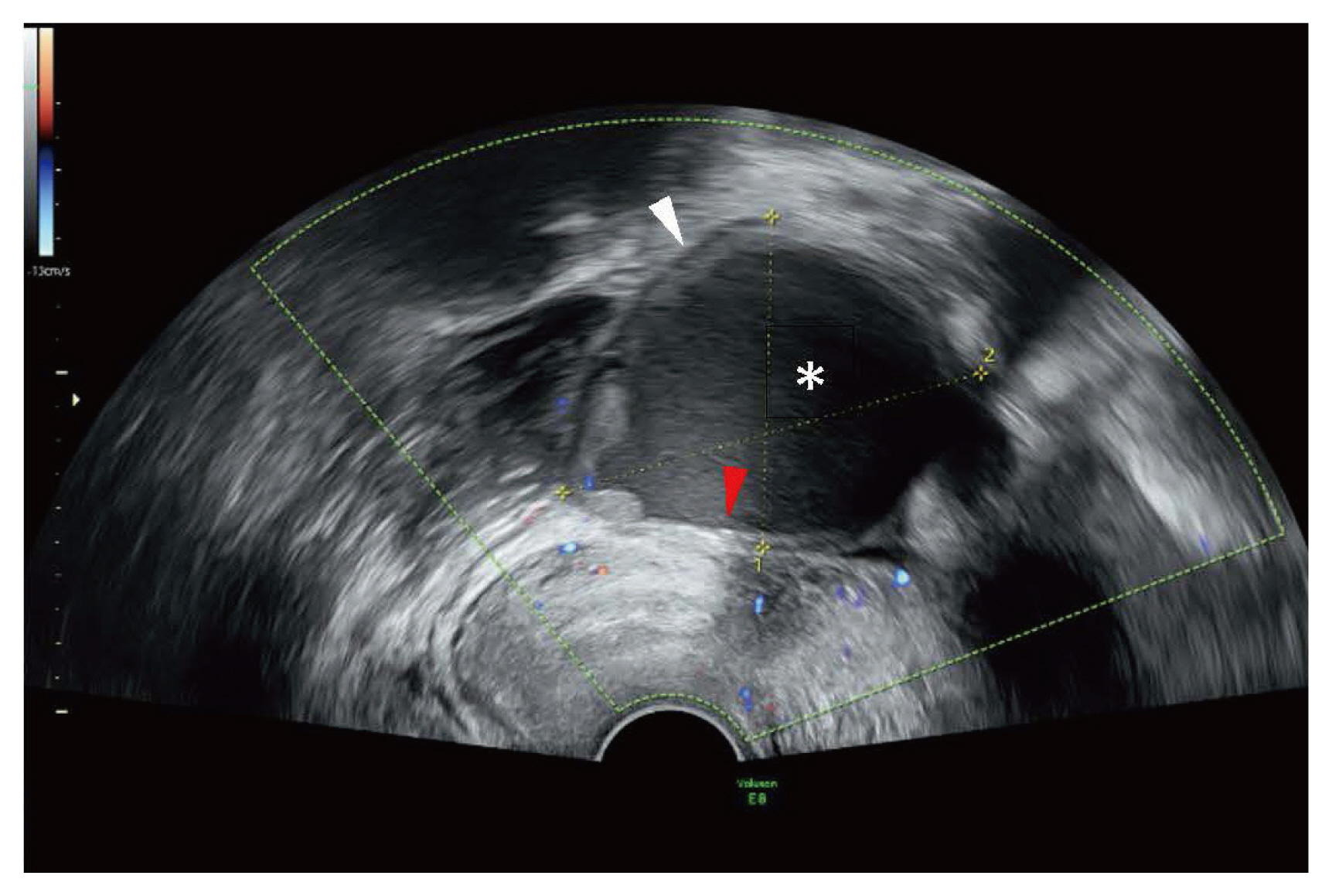
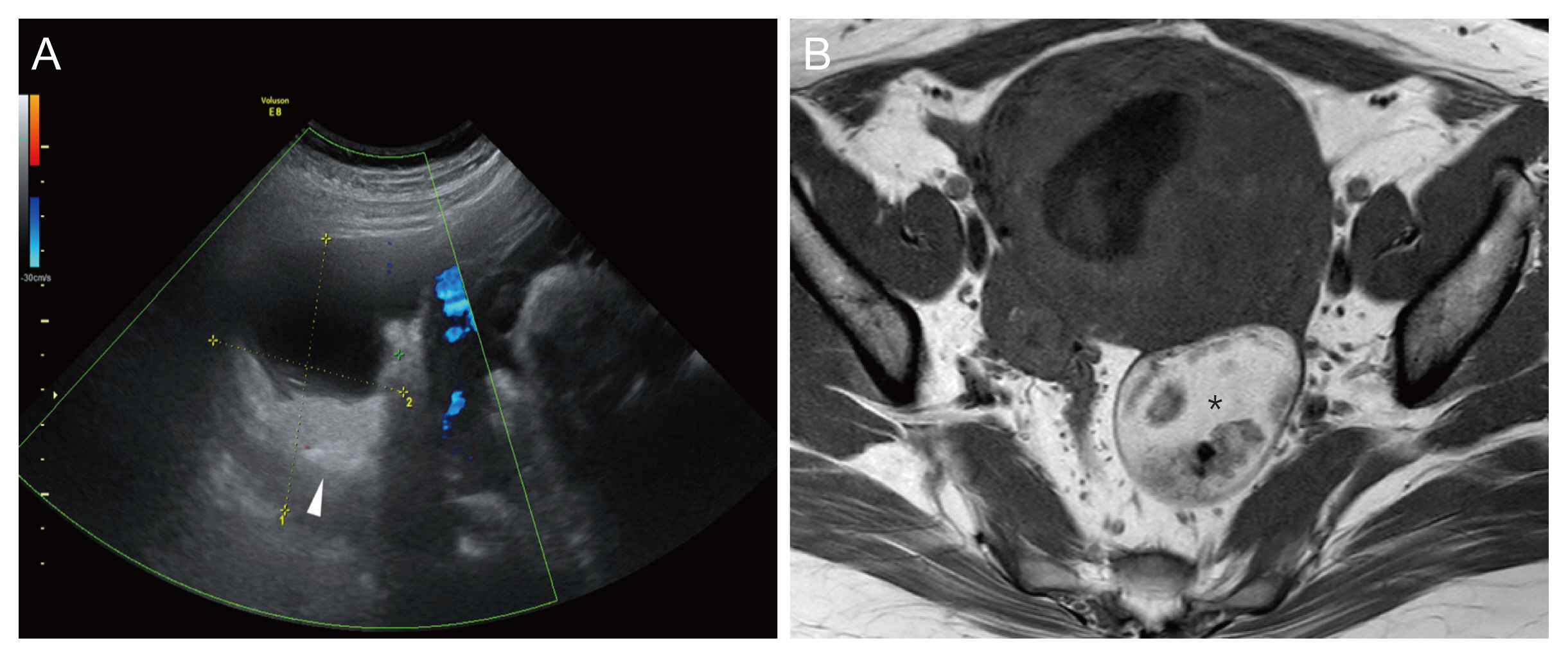
The appearance of endometriomas on US varies, including cysts and solid masses, which are affected by recurrent hemorrhage and degradation of blood clots [43]. Thick hypovascular walls and internal homogenous low echogenicity are the most common features (Fig. 1). Multiple cysts are common and show fluid-fluid levels in some cases. US also shows combined non-shadowing hyperechoic wall foci and multilocular lesions without neoplastic features [44]. Endometriomas may contain large calcifications that cause acoustic shadowing [45], and calcifications present as mural linear or punctate types. Non-vascular intracystic nodules may exist, which should be distinguished from the solid portion, with vascularity, in malignant masses. In addition, these cysts may have reticular patterns due to acute hemorrhage, which can be misinterpreted as hemorrhagic cysts [44]. During pregnancy, endometriomas may change into more homogenous, less fibrotic, and less definite nodules with band-like echoes on US [46]. Although reversible, these changes have been reported in up to 12% of pregnant women [47,48]. MRI might be helpful in diagnosing endometrioma and in excluding malignancy. On T1WI, endometriomas present as high signal intensity ovarian masses due to the high iron concentration from repetitive hemorrhage compared to whole blood. In contrast, due to hemosiderin in T2WI, endometriomas show low signal intensity, known as “shading.” This characteristic difference between T1WI and T2WI provides sensitivity and specificity of at least 90% for diagnosing endometriomas using MRI [49]. Low-signal-spiculated bands can be observed on T1WI and T2WI, and detection can be improved using fat-saturated sequences.
Because of the pregestational effects during pregnancy, occasionally, the wall of an endometrioma may decidualize and become a solid vascularized wall component with increased papillary intraluminal projections [50]. In such cases, distinguishing between vascularized malignant tumors and benign endometriomas may be difficult. Since both present with increased vascular flow, color Doppler is also not useful [51]. MRI findings may facilitate the differential diagnosis but are not conclusive. Additionally, cancer antigen-125 levels are not helpful in distinguishing between malignancy and decidualized endometrioma since they are generally elevated in normal pregnancies [52]. Owing to these limitations, patients tend to undergo surgery without a confirmed malignancy, and the diagnosis is only confirmed by the final pathology. Unfortunately, little is known about the proportion of endometriomas that undergo decidualization during pregnancy. Therefore, conservative management should be considered before performing invasive surgery.
6. Mature cystic teratoma
Mature cystic teratomas or dermoid cysts are the most common benign adnexal cysts, diagnosed in 20-40% of pregnant women with ovarian masses [53-55]. To date, no definite morphological changes induced by pregnancy have been reported. These cysts are bilaterally detected in up to 15% of the general population [56,57]. Malignant transformation is rare, accounting for 2% of all cases [58]. Because of the limited space due to the enlarged uterus, twisting of the teratoma on its pedicle can occur, but torsion is rarely reported [59]. Conservative management can be considered in pregnant women with a low risk of either malignancy or acute complications of the teratoma.
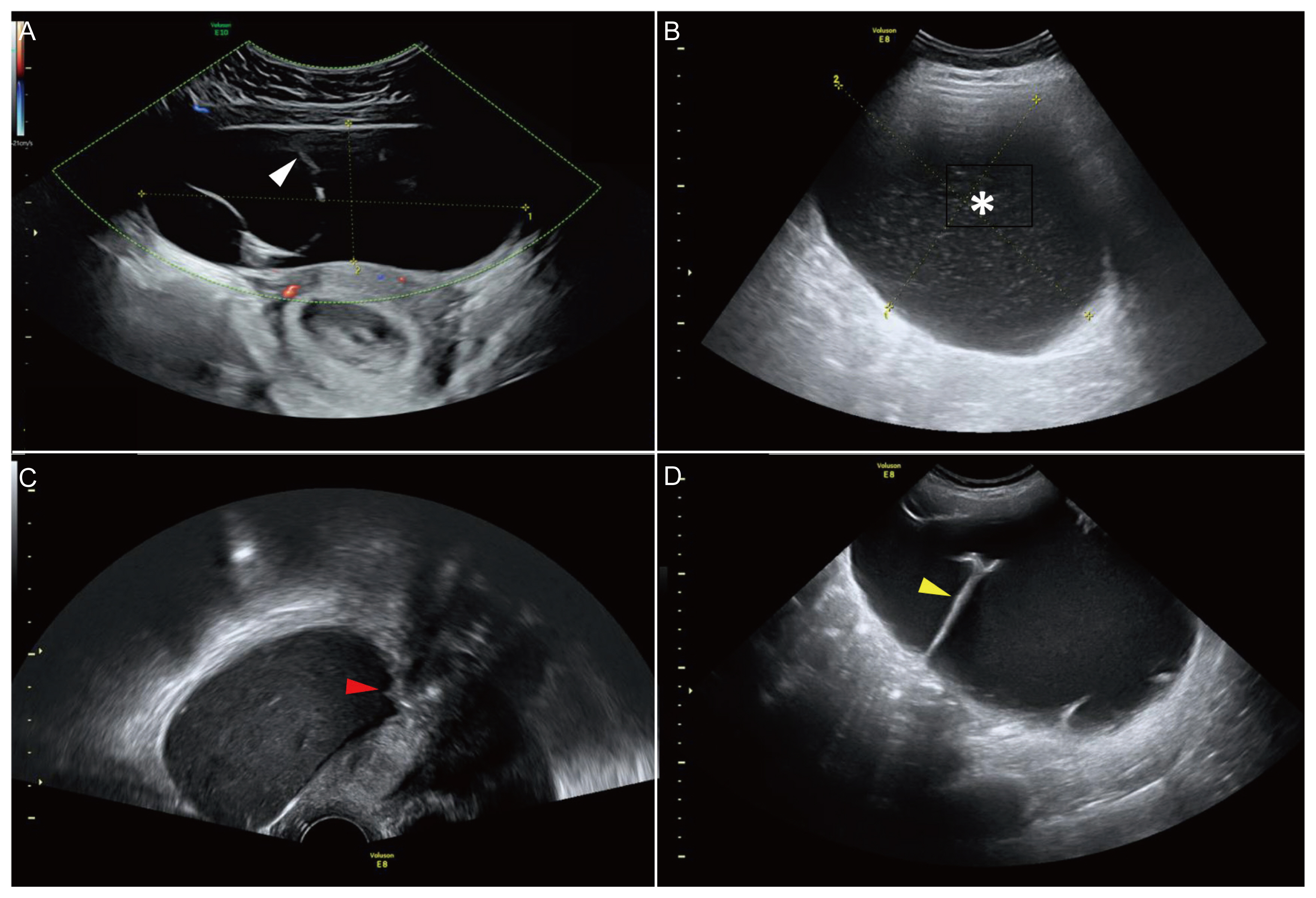
It has been reported that 95% of mature cystic teratomas are expected to be diagnosed using US [60] and there are three prominent common characteristics. First, hyperechoic nodules with acoustic shadowing on a hypoechoic background, known as Rokitansky nodules or “dermoid plugs” are one of the characteristic features of mature cystic teratomas (Fig. 2). A mixture of hair and sebum contributed to this appearance. Second, attenuation behind a large echogenic lesion, known as the “iceberg phenomenon, obscures the posterior wall of the cyst.” Third, multiple interdigitating lines and dots, representing floating hairs in the sebum, known as “dermoid mesh.” It has been reported that an adnexal mass with two or more characteristic sonographic features have a positive predictive value of 100% [61]. Occasionally, focal calcifications, which were present as internal heterogeneities, were observed. Fluid-fluid level is usually present, and the non-dependent or floating layer appears hyperechoic but hypoechoic in the dependent layer. In rare cases, multiple floating echogenic spherules and balls appear in the cyst [62].
On MRI, sebaceous and fat components show high signal intensity on T1WI; suppression of high signal intensity on T1WI with frequency-selective fat saturation indicates sebaceous and fat components and differentiates blood components that are not suppressed. In addition, chemical shift artifacts can be used to detect fat components and distinguish them from blood components in the frequency encoding direction [63].
7. Cystadenoma
In pregnant women, the most common types of ovarian cysts are epithelial serous or mucinous neoplasms, which are generally benign and referred to as cystadenomas. Cystadenomas account for up to 50% of benign ovarian neoplasms in the general population [31], with the serous subtype being the most common. Surgical intervention should be postponed if there are no definite suspicious signs of malignancy or emergent situations such as torsion, rupture, or obstruction of labor.
Serous cystadenomas present bilaterally in approximately 20% of cases. On US, it appears as a smooth-walled unilocular cyst that is difficult to distinguish from a functional cyst (Fig. 3). However, unlike functional cysts, serous cystadenomas are generally larger and do not spontaneously resolve during pregnancy. Therefore, simple cysts should be monitored during pregnancy to check for changes in size and characteristics. With significant changes, surgical resection should be considered [60]. Serous cystadenomas are approximately 10 cm on average but can measure up to 30 cm. Papillary projections may be present inside the cyst but are usually small or inapparent. Multiple thin hypoechoic septations were identified. Doppler imaging showed a high-resistance flow of the pulse wave. On T1WI, the contents of serous cystadenoma usually appear as a low signal intensity but sometimes as a high signal intensity. However, on T2WI, the contents of the cyst show high signal intensity; papillary projections present as high signal intensity lesions with low signal intensity in thin fibrous walls. If papillary projections inside the cyst appear as prominent small nodular protrusions, borderline tumors should be considered.
Bilaterality is scarce in mucinous cystadenomas, occurring in only 2-5% of cases [31,63], and suspicion of a borderline or malignant mucinous tumor should be considered. Papillary projections or solid components inside the cyst can also indicate borderline or malignant tumors. On US, one would typically find a large multiloculated cyst with multiple septations and hypoechoic locules. Pulsed-wave Doppler demonstrates high-resistance waveforms, resistive, and pulsatile indexes [63]. However, US may not be able to distinguish between mucinous and serous cystadenomas. On T1WI, the mucinous cystadenoma contents show low signal intensity, and depending on the concentration of mucinous components or hemorrhages, the locules show a higher signal intensity. On T2WI, the cyst contents showed high signal intensity, and the cyst walls were enhanced with gadolinium on contrast-enhanced magnetic resonance (CEMR). In most cases, US is sufficient to characterize mucinous tumors; however, MRI may be helpful if the sonographic findings are equivocal or unable to exclude malignancy. As mucinous cystadenomas are generally large, surgery should be considered to prevent torsion and malignancy if indicated.
8. Sex cord-stromal tumor
Although malignant sex cord-stromal tumors (usually granulosa cell tumors [GCT]) are more common than benign tumors, such as fibromas or fibrothecomas, they rarely complicate pregnancy and typically present as stage I, with a high cure rate when managed by surgery alone. Similar to cystadenomas, surgery should be avoided if there is little suspicion of malignancy or acute or clinical symptoms.
Ovarian fibromas are mostly unilateral and may be associated with Meigs syndrome in 1% of the patients [64]. On US, fibromas characteristically appear as solid hypoechoic lesions with edge shadows and acoustic shadowing, similar to fibroids, and calcifications with different degrees of vascularity are observed. Similar to uterine fibroids, fibromas predominantly show low signal intensity on T2WI, which is characteristic of fibrocollagenous stroma. Central cystic lesions may occur due to degeneration and edema in larger lesions. On T1WI, a low to intermediate signal intensity is observed. Different levels of enhancement are observed on CEMR. Calcified lesions present as low-signal foci on T1WI and T2WI. Degeneration may present with mixed features on both US and MRI.
GCTs are usually large and present as solid multilocular masses on US. Most GCTs appear as solid tumors with heterogeneous echogenicity. Hemorrhagic components are common inside cysts, and vascularity is increased on colored-power Doppler US. According to a study on the characteristics of MRI for GCT, varying signal intensities were observed on both T1WI and T2WI [65]. On T1WI, the GCT showed low to mixed intensity signals, whereas other sex cord-stromal tumors presented as hypointense or isointense masses. In contrast, on T2WI, GCT showed high or mixed intensity signals, which was different from other sex cord-stromal tumors that present as isointense and hyperintense masses. The study also reported that the average apparent diffusion coefficient value in the GCT group was lower than that in the other sex cord-stromal cell tumor group.
9. Paratubal cyst
Paratubal cysts are simple cysts that do not arise from the ovaries, but attach to the fallopian tube, broad ligament, or mesosalpinx. Because they are mostly benign and have no significant clinical implications, conservative management is usually warranted.
On US, paratubal cysts presented as simple locular cysts distant from the nearby ovary (Supplement Fig. 2). Their sizes vary, and although some are large, most are <1 cm. Paratubal cysts exhibit anechoic or hypoechoic echogenicity, their walls are largely inapparent, and they generally do not have septations, solid portions, or vegetation.
On MRI with T1WI, paratubal cysts have low signal intensity, and caution is warranted if high signal intensity is visible within cysts because it suggests hemorrhage due to torsion. In contrast, paratubal cysts show high signal intensity on T2WI, and heterogeneity inside the cysts suggests hemorrhage due to torsion.
10. Hydrosalpinx
A hydrosalpinx shows a “beads-on-a-string” sign due to hyperechoic mural nodules on US, which measure 2-3 mm on the cross-section and distended fluid-filled tubal structures. This sign represents flattened and fibrotic intratubal fluid folds induced by progressive fluid accumulation and distention due to a non-patent tube. In some chronic cases, the tube wall is thickened (>5 mm) [63]. Identification of the normal ipsilateral ovary may help diagnose hydrosalpinx [66]. If suspicious features are present, including solid elements or papillary projections inside the tubular cystic lesion, fallopian tubal carcinoma should be considered, which is rare [67]. Surgery is generally indicated in cases of acute abdominal pain that does not regress.
MRI is helpful in identifying the ipsilateral ovary as separate from the suspected hydrosalpinx. A fluid-filled tubular structure is visible on T2WI. Signal intensity varies in T1WI, depending on the contents of the tube; simple fluid presents as low signal intensity, but fluid with diverse protein components presents as intermediate to high. Coronal views best demonstrated a hydrosalpinx.
11. Uterine fibroid
According to one study, fibroids are common, especially uterine fibroids, and occur in up to 10% of pregnant women. Uterine fibroids occasionally present as pedunculations. The characteristic appearance remains unchanged during pregnancy, but its size may increase owing to hormonal effects. Occasionally, patients present with acute pain due to internal degeneration, hemorrhage, or torsion. Conservative management is possible if patients are asymptomatic and malignant potential can be ruled out.
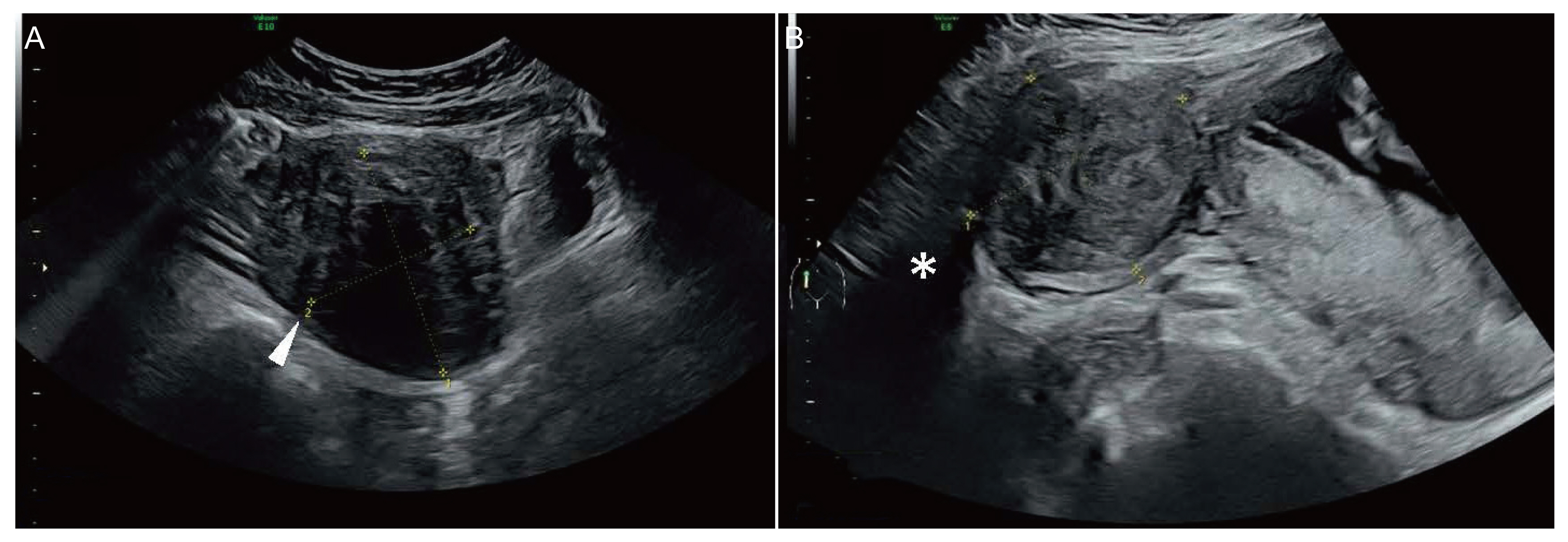
On US, uterine fibroids show a hypoechoic solid mass with edge refraction and posterior acoustic shadowing (Fig. 4). Cystic and calcified components may exist in cysts owing to internal degeneration and necrosis. Pedunculated fibroids generally present with feeding vessels that are connected to the uterus. The torsion of a pedunculated fibroid shows a variable lack of vascularity on color Doppler imaging.
MRI may be helpful in demonstrating the characteristic low signal intensity on T2WI, which is associated with a fibrocollagenous stroma. Larger fibroids lacking vascular supply might undergo internal cystic or hemorrhagic degeneration, which might result in mixed features on both US and MRI.
12. Borderline ovarian neoplasm (low malignant potential)
Most epithelial ovarian neoplasms are borderline tumors, also known as tumors with low malignant potential, showing good prognosis and benign courses, and malignancy is rare. These neoplasms are often present in women of childbearing age, and one-third of cases are diagnosed in women aged ≤40 years [68,69]. It has been reported that 2.15-13.5% of all adnexal masses detected during pregnancy consist of borderline ovarian neoplasm and ovarian cancers [70]. Fertility-sparing surgery is one of the treatment options, but it may result in a higher risk of recurrence, which occurs in 45-56% of patients in advanced stages [71-75]. There is currently no consensus on the standard management of borderline ovarian neoplasms; thus, conservative management is typically recommended unless malignant features are observed.
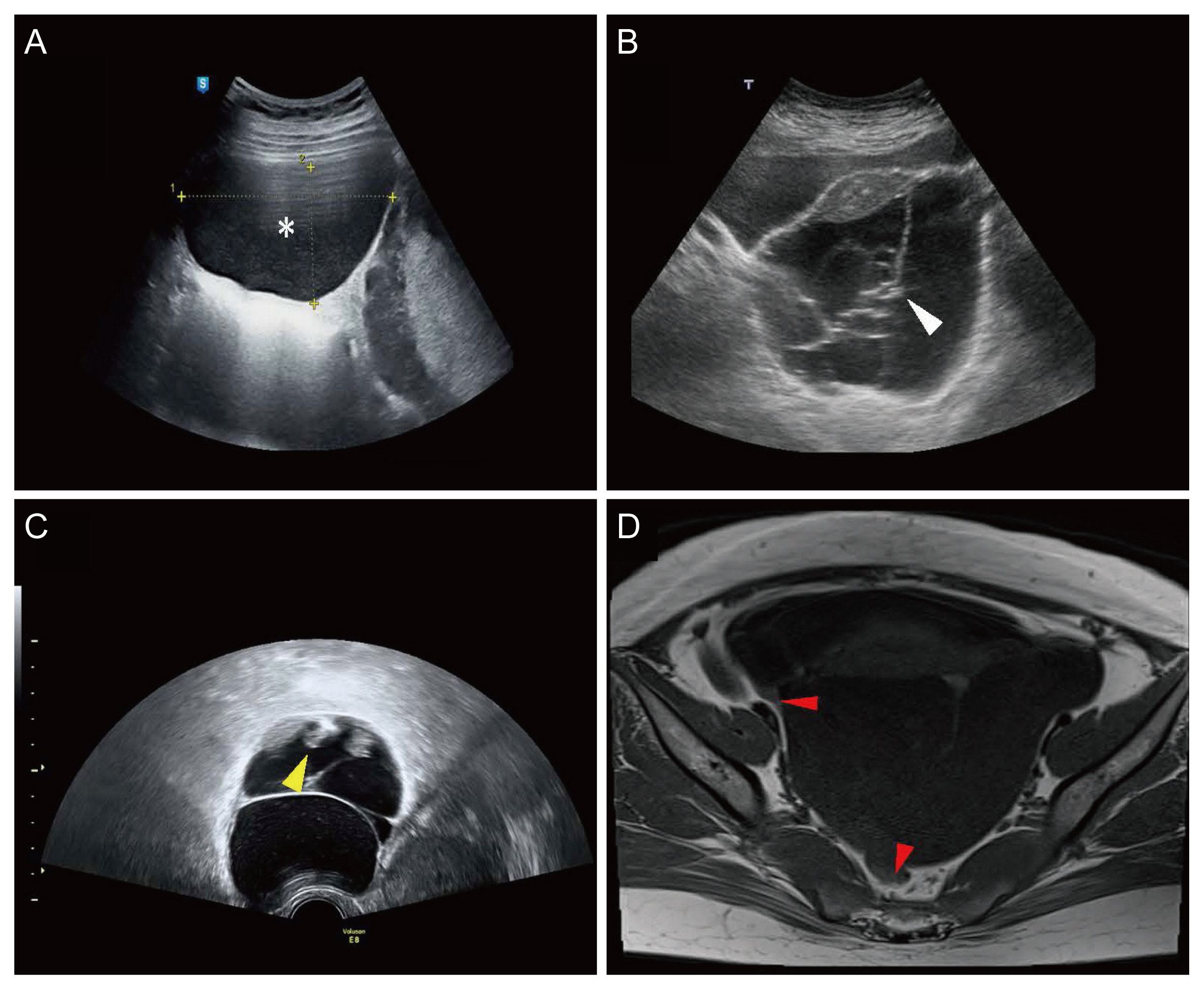
Sonographic features suggestive of borderline tumors were as follows: 1) a unilocular cyst with an ovarian crescent sign (which represents the rim of the normal ovarian tissue adjacent to the lesion), 2) multiple vascular mural wall nodules, 3) papillary projections, and 4) a cystic lesion associated with a well-defined multilocular nodule with a “honeycomb” appearance (Fig. 5). The ovarian crescent sign might help to exclude invasive ovarian cancer in some cases [31]. A previous study confirmed that US is feasible for characterizing borderline ovarian neoplasms and their recurrence [76].
The international ovarian tumor analysis describes the following characteristic US features that can be helpful in distinguishing between benign and malignant masses. The following are characteristic findings of malignant masses: irregular solid tumor, multilocular and irregular mass >10 cm, presence of three or more papillary projections, vascular pattern, ascites, and/or metastases [77,78]. However, their efficacy in pregnant women with adnexal masses has not been reported.
Borderline tumors generally demonstrate one or more findings suggestive of malignancy on MRI [79], including bilaterality, tumor size >4 cm, predominantly solid mass, cystic tumors with projections, or contrast enhancement [80]. Additionally, several types of borderline tumors with different MRI findings have been reported in the literature. For example, a unilocular cyst with papillary projections from the cyst wall may be observed; in particular, it presents with high and low signal intensity papillary projections from the cystic wall on T2WI. Minimally septate cysts with papillary projections involving the wall and septa. On T1WI, fat-saturated MRI shows enhancing papillary projections inside the cysts, in contrast to the low signal intensity on T2WI. Markedly septate cysts with plaque-like excrescences, especially for the mucinous subtype, are clearly visible on T1WI and fat-saturated MRI. Additionally, borderline tumors may present as predominant solid masses with exophytic projections on MRI, particularly in the serous subtype. Exophytic projections may resemble the external branching of cauliflowers, and T1WI fat-saturated MRI shows enhancement involving cyst walls and projections inside the cysts.
13. Germ cell tumor
Dysgerminoma is a germ cell tumor that accounts for up to 5% of ovarian malignancies. It is the most common malignancy diagnosed during pregnancy (up to 15-20%), excluding epithelial ovarian neoplasms with low malignancy potential [63,81,82]. Other germ cell tumors, such as embryonal carcinomas, immature teratomas, and yolk sac tumors, rarely occur during pregnancy. As dysgerminoma has a good overall prognosis, the continuation of pregnancy after the staging is generally accepted. Surgery should be performed in the second trimester because of the decreased risk of miscarriage and the size of the uterus, which allows access during this time [83-87]. Surgical exploration during the third trimester has also been reported to be associated with premature labor [88,89].
On US, a dysgerminoma presents as a multilobulated solid ovarian mass with prominent fibrovascular septations and calcifications. It typically presents heterogeneous echogenicity but may contain anechoic or hypoechoic areas because of internal hemorrhage or necrosis. A low signal intensity mass is seen on T1W1, while a mass with intermediate signal intensity with low signal intensity septations and high signal intensity necrotic areas are seen on T2W1. On CEMR, fibrovascular septations were homogenously enhanced. In addition to US and MRI, elevated levels of α-fetoprotein and lactate dehydrogenase can also help in the diagnosis since these markers are frequently reported in dysgerminoma during pregnancy [90-92].
14. Malignant epithelial ovarian neoplasm
Benign mucinous and serous epithelial ovarian neoplasms are commonly detected during pregnancy. Mucinous and serous cystadenocarcinomas may also occur. On US, they present as multilocular cystic masses and may also exhibit irregular cyst walls, papillary projections, thick septations, papillary projections, and vascular mural or septal nodules. Because mucinous and serous cystadenocarcinomas have similar US findings, MRI may help differentiate them. Malignancy should be suspected if the papillary projections or septations are extensive. In cases of suspected malignancy, a thorough inspection is warranted, including the presence of ascites, peritoneal seeding, or omental cakes. In patients who desire to maintain their current pregnancy, only cystectomy or adnexectomy should be performed because complete cytoreduction is impossible during pregnancy. Platinum-based chemotherapy and cytoreductive surgery should be administered to patients after delivery. The optimal time to start chemotherapy while avoiding fetal toxicity is at the end of the 14th gestational week, which is considered the end of the first trimester [93].
On US, a serous cystadenocarcinoma presents as a cystic adnexal mass with heterogeneous echogenicity. Thick walls, multiple septations, papillary projections, or nodules can also be observed. On MRI, a cystic mass with low-to-intermediate intensity was observed on T1WI. However, T2WI showed a high intensity cystic mass with solid portions of heterogeneous intensity. CEMR shows the solid components better. Mucinous cystadenocarcinomas are less common than serous cystadenocarcinomas and present as multiloculated cystic masses with solid components. On US, a multiloculated cystic mass with heterogeneous echogenic patterns and solid portions was demonstrated. On MRI, signal intensity may vary depending on the amount and concentration of mucin. A loculus containing watery mucin can present a low signal intensity compared with thick mucin on T1WI. The signal intensity differs between cysts with watery and thick mucin on T2WI; watery mucin shows high signal intensity, but thick mucin shows low signal intensity.
Conclusion
Adnexal masses detected during pregnancy are mostly benign tumors, which are also observed in the non-pregnant population, and malignant tumors such as dysgerminoma are scarce. Certain unique adnexal masses are exclusively visible during pregnancy, such as hyperreactio luteinalis, theca lutein cysts, and luteoma. Differential diagnosis of adnexal masses in pregnant women is important because an incorrect diagnosis can result in inappropriate treatment and affect perinatal outcomes. US can be helpful in the differential diagnosis of adnexal masses based on the characteristic findings in pregnant women. Similarly, MRI can assist with characteristic T1WI and T2WI findings, where US findings are indeterminate or equivocal. Therefore, with the use of US and MRI, an accurate diagnosis with appropriate treatment is possible. Important key US and MRI findings of adnexal masses during pregnancy are summarized in Tables 1, 2, respectively. If malignancy is not definite, conservative management should be considered first; however, prompt surgical intervention or chemotherapy should be considered when malignancy is highly indicated by the presence of characteristic features or is pathologically confirmed.


")


