


Introduction
Vitamin D is known to have a major influence on bone health through regulation of calcium phosphorus homeostasis. Recently, the female reproductive system has been defined as one of the non-classical target organs of vitamin D. Vitamin D receptor expression was identified in ovarian granulosa cells as well as other female reproductive organs, including endometrium and the uterus. These findings suggest that vitamin D may have a potential role in female reproduction [1-3]. Several studies have demonstrated a direct effect of vitamin D on ovarian folliculogenesis and steroidogenesis in animal and human cell-line studies; vitamin D receptor null mutant mice have impaired folliculogenesis and vitamin D stimulated steroidogenesis in human ovarian cells [4,5].
Anti-Müllerian hormone (AMH) is produced by granulosa cells of small antral and pre-antral follicles, and along with antral follicle counts (AFCs), is a well-known representative marker for ovarian reserve. AMH plays a crucial role in folliculogenesis and is not affected by the menstrual cycle, making it the most widely used ovarian marker. Although it is still unclear how vitamin D affects female reproductive function, interest in a potential relationship between vitamin D and ovarian reserve markers, particularly AMH, has increased since reports on the effect of vitamin D on follicular development have accumulated. Moreover, the presence of a functional vitamin D response element in the AMH gene promotor region was demonstrated [6] and experimental results in animal and human granulosa cells have also reported that vitamin D affects AMH signaling [7,8]. These basic research studies have indicated that vitamin D deficiency may alter gonadal function through abnormal AMH signaling. However, discrepancy exists in clinical study findings under various settings, where some suggest a significant association between vitamin D levels and ovarian reserve, while others have not found any correlation between vitamin D and ovarian reserve markers [9,10]. Considering these contradictory results, we performed this prospective observational study to examine serum 25-hydroxyvitamin D [25(OH)D] levels in patients with secondary amenorrhea (SA) and investigate the relationship between serum 25(OH)D levels and ovarian reserve markers (i.e., serum AMH and AFC) in these women.
Materials and methods
1. Study design and participants
This study was conducted as a prospective cohort study for 12 months from March 2018 to February 2019. The study population was comprised of patients evaluated by a single reproductive endocrinologist for symptoms of SA. A total of 78 participants were initially recruited to the study. Three participants refused to participate in the study and 12 participants were excluded due to the study’s exclusion criteria. The exclusion criteria of this study were as follows: 1) women who had taken hormonal medication including oral contraceptives or vitamin D supplements within the previous 6 months; 2) women who had ovarian surgery, chemotherapy or radiotherapy; 3) women who took medication and/or were diagnosed with a systemic disease that can affect menstruation (e.g., psychoaffective medicine, thyroid hormone, diabetes, hyperprolactinemia); 4) women who refused to participate this study. Patients’ information including age, parity, height, body weight, cause of SA, season of blood sampling (i.e., to account for seasonal changes in vitamin D), current medications and other past medical history were recorded. Gynecologic ultrasonography was also performed on their first study visit. To determine cause of SA, polycystic ovarian syndrome (PCOS) was diagnosed on the basis of Rotterdam criteria [11], and primary ovarian insufficiency (POI) was regarded as 2 recordings of serum follicular stimulating hormone (FSH) levels of more than 40 IU/L at least one month apart in a woman aged under 40 years (i.e., >2 standard deviation [SD] less than the mean menopausal age) [12]. All patients whose etiology of SA was not determined, excluding women with POI and PCOS, were deemed unexplained chronic anovulation [13]. PCOS in adolescents was only diagnosed in women with at least 2 years after menarche and who had SA, hyperandrogenemia, and increased ovarian volume on ultrasound [14].
Venous blood samples of all participants were taken for the measurement of serum 25(OH)D and AMH on the same day, prior to treatment for SA. Vitamin D deficiency was defined as serum 25(OH)D levels <20 ng/mL based on the Endocrine Society clinical practice guidelines [15].
2. Serum vitamin D, hormones and ovarian reserve markers measurements
1) Serum 25-hydroxyvitamin D
Each participant’s venous sample was drawn into a serum separation tube (SST) and serum 25(OH)D was measured using a DIA source 25 OH Vitamin D3 radioimmunoassay kit (DIAsource ImmunoAssays S.A., Louvain-La-Neuve, Belgium) with immunoassay device (gamma 5) and presented in ng/mL. The total imprecision coefficient of variance was 5.38% at a concentration level of 6.35 ng/mL and 4.96% at 37.71 ng/mL.
2) Serum follicular stimulating hormone
Each participant’s venous sample was drawn into an SST and serum FSH was measured using an Elecsys FSH electrochemiluminescence immunoassay kit (Roche Diagnostics GmbH, Mannheim, Germany) with immunoassay device (Cobas e 801) and presented in mIU/mL. The total imprecision coefficient of variance was 3.1% at a concentration level of 48.8 mIU/mL and 2.3% at 17.4 mIU/mL.
3) Anti-Müllerian hormone and antral follicle counts
Blood drawn into the SST was centrifuged within 1 hour (3,000 rpm for 10 minutes). The separated serum was measured for AMH level by ECLIA method with Elecsys AMH kit (Roche Diagnostics GmbH) on a Cobas e 601 immunoassay analyzer and presented in ng/mL. The total imprecision coefficient of variance was 3.5% at a concentration level of 0.042 ng/mL and 3.4% at 0.20 ng/mL. An AFC was measured by one gynecologist on the participant’s first visit in all individuals with gynecologic ultrasonography. AFC represented the total number of all antral follicles, from 2 mm to 10 mm in size, in both ovaries.
3. Statistical analysis
Categorical variable data are presented as a frequency with percentage and continuous variables are presented as a group mean±SD. Differences in study participants’ characteristics were compared across subgroups with a χ2 test or Fisher’s exact test for categorical variables and independent t-test or Mann-Whitney’s U test for continuous variables, as appropriate. An analysis of variance with Duncan’s post hoc test or Kruskal-Wallis test with Dunn’s post hoc test were also employed, as appropriate. To check if data were normally distributed, we used a Shapiro-Wilk test. Partial correlation coefficients controlling for age and body mass index (BMI) were estimated to investigate the linear relationship between 2 continuous variables. Univariate linear regression analysis between each variable (age, BMI, vitamin D, season), and multivariate linear regression analysis with all covariates, were performed to identify the regression coefficients for factors related to the log10 AMH, AFC, PCOS and POI. For data visualization, box plot and scatter plot were also displayed. Finally, logistic regression analysis was performed to identify whether vitamin D deficiency is associated with PCOS as a cause of SA. All statistical analyses were carried out using SPSS 24.0 (SPSS Statistics for Windows 24.0; IBM Corp., Armonk, NY, USA) statistical software and P-values less than 0.05 was considered statistically significant.
Results
Sixty-three participants with SA were finally included in this study. The mean age of participants was 26.11±8.05 years and the mean BMI was 22.75±4.75 kg/m2. Participants’ baseline and biochemical characteristics of patients are shown in Table 1. Of all participants, 42.9% (n=27) were vitamin D deficient (<20 ng/mL) and 57.1% (n=36) had normal vitamin D levels (≥20 ng/mL). The mean AMH levels and AFC were 10.86±8.94 µ/L and 15.23±7.65 in the vitamin D deficient group, and 7.24±5.62 µ/L and 12.30±6.95 in the normal vitamin D group, respectively. The difference in BMI, cause of amenorrhea, season of blood sample and FSH levels between the 2 groups did not reach statistical significance, but age was significantly higher in the normal vitamin D group.
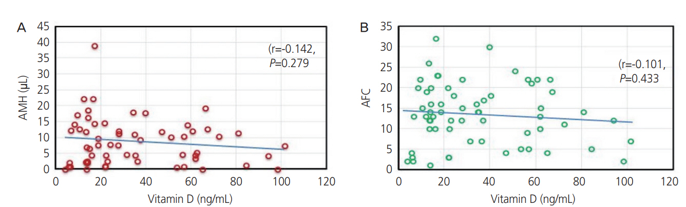
There was no correlation between serum vitamin D and AMH levels (r=−0.142, P=0.279) or AFC (r=−0.101, P=0.433) in all participants (Fig. 1). After univariate linear regression analysis of log10 transformed AMH or AFC with all covariates, multivariate linear regression analysis of log10 transformed AMH or AFC with vitamin D, age and BMI was performed with adjustment for confounding variables. While there was a significant association between serum AMH levels and age, there were no significant relationships between serum AMH levels or AFC and serum vitamin D levels: regression coefficient for vitamin D predicting log10 AMH: −0.002, standard error (SE): 0.004, P=0.557 (Table 2), regression coefficient for vitamin D predicting AFC: −0.042, SE: 0.040, P=0.304 (Table 3). In the univariate and multivariate linear regression of PCOS modelled with vitamin D levels, there was also no significant association between PCOS and serum vitamin D evels (Table 4). The results of univariate and multivariate linear regression of POI modelled with vitamin D levels was also similar: regression coefficient±SE: 0.001±0.002, P=0.641, −0.001±0.002, P=0.557, respectively). However, serum vitamin D levels for each SA cause group demonstrated lower tendency for vitamin D levels in the PCOS or POI groups than in the unexplained chronic anovulation group (31.07±22.05, 31.37±31.89, 48.92±27.69, respectively, P=0.056) (Fig. 2). In the logistic regression analysis, women with vitamin D deficiency had an increased chance of having PCOS as cause of SA than those with normal vitamin D levels (adjusted odds ratio [OR], 7.559; 95% confidence interval, 1.28-44.65; P=0.026) after adjusting for BMI, age and seasonal variation of vitamin D.
Discussion
Our study demonstrated that serum 25(OH)D levels were not related with ovarian reserve markers among SA patients, even after adjustment for age, BMI, cause of amenorrhea, and seasonal variation of vitamin D. However, mean serum vitamin D levels were different depending on the cause of amenorrhea, however the difference did not reach statistical significance (Fig. 2). In particular, vitamin D levels in the participants with PCOS or POI demonstrated a tendency towards lower serum vitamin D levels than those in participants with unexplained chronic anovulation. In addition, women with vitamin D deficiency had an increased chance of having PCOS than those with normal vitamin D levels, after adjustment for clinical factors by logistic regression model. We did not calculate the adjusted OR for POI since participants with POI were a very small portion of the study participants. The lack of correlation between serum vitamin D levels and ovarian reserve markers across all participants in our study is in agreement with several recent studies that also demonstrated no correlation. However, the result of lower vitamin D levels in participants with PCOS in this study needs to be taken with caution, considering the heterogeneous results of previous studies [16,17]. To date, the role of low vitamin D levels on AMH regulation or the pathophysiology of PCOS is unclear, with some previous reports suggesting that this vitamin D deficiency is likely due to obesity or a metabolic syndrome that is frequently associated with PCOS [18,19].
While basic research has reported that vitamin D affects ovarian folliculogenesis and steroidogenesis, there is limited clinical information on how vitamin D affects ovarian markers such as AMH. Wojtusik et al. [9] have reported a dosedependent decrease in AMH mRNA levels in granulosa cells after treatment with vitamin D. In another recent study, human granulosa cells treated with vitamin D exhibited altered AMH signaling and they demonstrated an inverse correlation between vitamin D status in follicular fluid and AMH receptor-II (AMHT-II) mRNA gene expression [8]. Binding AMH to AMH receptor-II (AMHR-II) is known to suppress follicular maturation, by inhibiting primordial follicle recruitment into the growing follicle pool, and by decreasing the sensitivity of follicles to FSH [10]. Since vitamin D had an effect on downregulation of AMHR-II gene expression, the phosphorylation process, and nuclear localization after AMH-AMHR-II binding [8], it may be involved in promoting follicle development by altering AMH production pattern and FSH sensitivity in ovarian granulosa cell [20,21].
Previous studies on the relationship between vitamin D and ovarian reserve markers, particularly AMH, have had inconsistent results. For instance, Merhi et al. [22] found a positive relationship between serum vitamin D and AMH levels and suggested vitamin D deficiency might be associated with lower ovarian reserve in late reproductive aged women. Dennis et al. [23] also suggested that vitamin D might have a positive effect on AMH production in adults, and the extent of seasonal variation in women’s AMH levels correlated with the extent of their variation in vitamin D levels. In another study of 1,430 premenopausal women, a negative correlation was found between serum vitamin D levels and urinary FSH levels, suggesting that low vitamin D levels may influence ovarian reserve and earlier menopause [24]. Meanwhile, a recent cross-sectional study that included 283 infertile women, revealed no significant association between vitamin D and ovarian reserve markers (AMH, AFC) [25], which is in agreement with a prospective study that failed to demonstrate any beneficial effect of vitamin D supplementation on normalization of serum AMH levels [26].
In this study, generally healthy women should be included as controls. However, it was difficult to recruit healthy women to volunteer to have their AMH and vitamin D levels measured, so we conducted this study in patients with SA who required AMH measurements for diagnosis. These SA patients included women with various levels of ovarian reserve, and it was assumed that unexplained chronic anovulatory patients were similar to healthy women with normal ovarian reserve. Indeed, participants with unexplained chronic anovulatory in our study demonstrated normal ovarian reserve (data not shown). In addition, cholecalciferol, the main natural source of vitamin D, is affected by sun exposure and outdoor activity, so participants’ duration of outdoor activity should also be measured. However, we did not conduct a survey of patients’ outdoor activity as subjective factors could have been reflected in this evaluation. Although the validity of AMH measurements in those younger than 20 years of age has not been established, we included participants in this age group and their AMH measurement, as the small number of girls in their late teens in this study were considered to have reached sufficient maturity.
This study is the first report on the relationship between serum vitamin D levels and ovarian reserve in Korean women. This study was possible because it was conducted in patients with SA, which included women with various levels of ovarian reserves. Furthermore, this study is strengthened by its prospective design, which has a low risk of confounding bias, the fact that both AFC and AMH were measured as a marker of ovarian reserve, and finally AMH was measured on the same day as 25(OH)D.
However, this study has several limitations. Firstly, this study has small study population to then analyze by subgroup(s) of amenorrhea causes, which increases the risk of selection bias or type II error. Secondly, healthy women with normal menstrual cycles, who were not SA patients, were not included as controls. Thirdly, this study was observational in nature, so we cannot interpret these results in terms of causality. Despite of these limitations, this study is the first prospective observational study of the relationship between vitamin D and ovarian reserve in Korean women with SA, suggesting no association between them. In this regard, it is not necessary to routinely measure serum vitamin D levels in SA patients with abnormal ovarian reserve. However, it would be meaningful to provide vitamin D supplementation, after measuring serum vitamin D concentration in PCOS patients, considering the low vitamin D levels in PCOS patients in this study. Nevertheless, given the uncertainty of how vitamin D affects female reproduction and the limitations of this study, no clear conclusion can be drawn. Further study is required on the relationship between vitamin D levels and obesity or metabolic abnormalities in patients with PCOS.
In conclusion, this observational study demonstrated that there was no correlation between serum vitamin D levels and ovarian reserve markers in SA patients, but that vitamin D deficiency may be linked to PCOS patients. However, given the limitations of this observational study, further prospective research with a larger population and age-matched control group should be investigated to draw more definite conclusions on the relationship between vitamin D levels and ovarian reserve markers.


")


